Daphna Link1, Netanell Avisdris1,2, Xingfeng Shao3, Liat Ben-Sira4,5, Leo Joskowicz2, Ilan Gull6, Danny J.J Wang3, and Dafna Ben-Bashat1,5
1Sagol Brain Institute, Tel Aviv Sourasky Medical Center, Tel Aviv, Israel, 2School of Computer Science and Engineering, The Hebrew University of Jerusalem, Jerusalem, Israel, 3Laboratory of FMRI Technology (LOFT), USC Stevens Neuroimaging and Informatics Institute, Keck School of Medicine, University of Southern California, Los Angeles, CA, United States, 4Division of Pediatric Radiology, Tel Aviv Sourasky Medical Center, Tel Aviv, Israel, 5Sackler Faculty of Medicine & Sagol School of Neuroscience, Tel Aviv University, Tel Aviv, Israel, 6Ultrasound Unit, Lis Maternity Hospital, Tel Aviv Sourasky Medical Center, Tel Aviv, Israel
1Sagol Brain Institute, Tel Aviv Sourasky Medical Center, Tel Aviv, Israel, 2School of Computer Science and Engineering, The Hebrew University of Jerusalem, Jerusalem, Israel, 3Laboratory of FMRI Technology (LOFT), USC Stevens Neuroimaging and Informatics Institute, Keck School of Medicine, University of Southern California, Los Angeles, CA, United States, 4Division of Pediatric Radiology, Tel Aviv Sourasky Medical Center, Tel Aviv, Israel, 5Sackler Faculty of Medicine & Sagol School of Neuroscience, Tel Aviv University, Tel Aviv, Israel, 6Ultrasound Unit, Lis Maternity Hospital, Tel Aviv Sourasky Medical Center, Tel Aviv, Israel
Placental functional-structural MRI assessment, with fetal brain
and body volumes at late gestation are presented. Increased normal placental
perfusion compared to earlier gestation, and differences in normal placental
and fetal characteristics vs. fetal growth restriction, are shown.
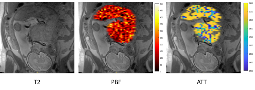
Figure 1: Representative Placental blood flow
(PBF) and Arterial Transit Time (ATT) results, for one placenta (GA = 32 weeks)
superimposed on the corresponding T2 anatomical image.
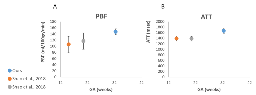
Figure 2: Mean Placental Blood Flow (PBF) (A)
and Arterial Transit Time (ATT) (B) values of three GA. The first two early GA
(mean of 15 weeks –orange, and 20.5 weeks- gray, are taken from5),
while the late GA (mean of 32.5 weeks, blue) demonstrates our results.