Bella Fadida-Specktor1, Dafna Ben Bashat2,3, Daphna Link Sourani2, Netanell Avisdris1,2, Elka Miller4, Liat Ben Sira3,5, and Leo Joskowicz1
1School of Computer Science and Engineering, The Hebrew University of Jerusalem, Haifa, Israel, 2Sagol Brain Institute, Tel Aviv Sourasky Medical Center, Tel Aviv, Israel, 3Sackler Faculty of Medicine & Sagol School of Neuroscience, Tel Aviv University, Tel Aviv, Israel, 4Medical Imaging, Children’s Hospital of Eastern Ontario, University of Ottawa, Ottawa, ON, Canada, 5Division of Pediatric Radiology, Tel Aviv Sourasky Medical Center, Tel Aviv, Israel
1School of Computer Science and Engineering, The Hebrew University of Jerusalem, Haifa, Israel, 2Sagol Brain Institute, Tel Aviv Sourasky Medical Center, Tel Aviv, Israel, 3Sackler Faculty of Medicine & Sagol School of Neuroscience, Tel Aviv University, Tel Aviv, Israel, 4Medical Imaging, Children’s Hospital of Eastern Ontario, University of Ottawa, Ottawa, ON, Canada, 5Division of Pediatric Radiology, Tel Aviv Sourasky Medical Center, Tel Aviv, Israel
We aimed to develop an automatic method for fetal total body segmentation from MRI data and to create a large dataset of normal fetuses. The method achieved high performance for two different sequences. Volumetric fetal body database was created and was in line with ultrasound growth chart.
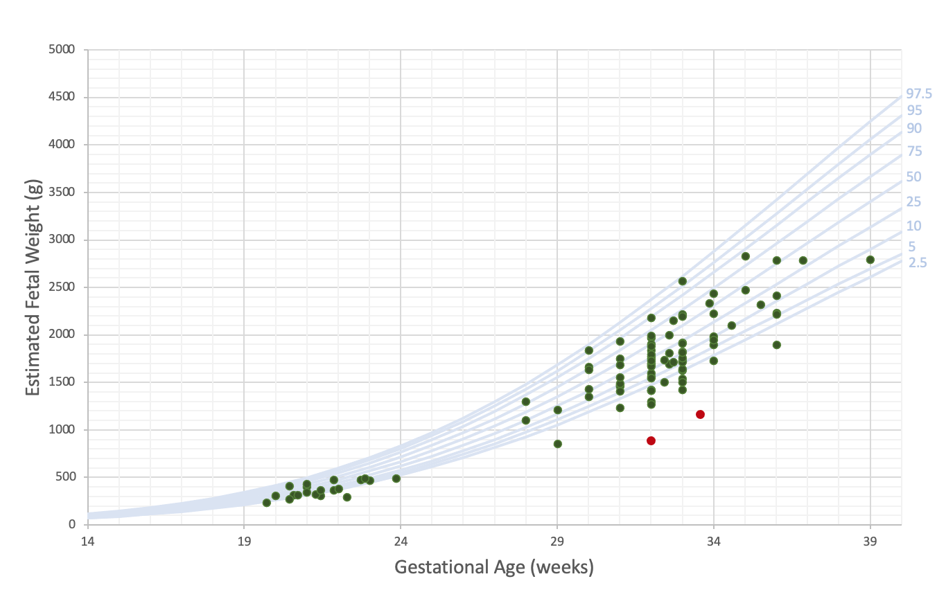
Fig. 2: Estimated fetal volume as a function of gestational age (green points) overlaid on ultrasound fetal body growth chart4 (blue curves representing percentiles). Red points correspond to IUGR fetuses.
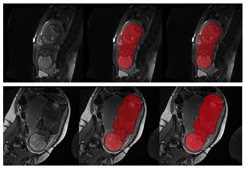
Fig. 1: Illustrative examples of the FIESTA (upper row) and TRUFI (lower row) fetal body segmentation results of two cases. Left: original image, middle: body segmentation result, right: manual ground truth.