Qian Cai1, Huan jun Wang1, Yi ping Huang1, Long Qian2, Yan Guo1, Zhi hua Wen1, Long yuan Ouyang1, and Mei qin Li1
1Department of Radiology, The First Affiliated Hospital of Sun Yat-sen University, Guangzhou, China, 2MR Research, GE Healthcare, Beijing, China
1Department of Radiology, The First Affiliated Hospital of Sun Yat-sen University, Guangzhou, China, 2MR Research, GE Healthcare, Beijing, China
Synthetic MRI are useful in noninvasively discriminating
low- and high-grade bladder cancer.
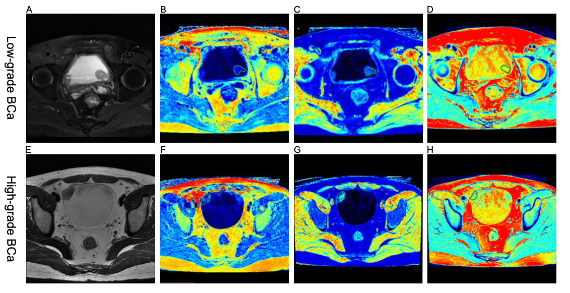
Figure 1 (A-D) A 58-year-old woman with low-grade BCa. (A) Axial T2-weighted image, (B-D) SyMRI-derived pseudo-colored maps (T1, T2, and PD, respectively) indicate that the mean T1, T2, and PD value are 27.27 msec, 26.18 msec, and 71.67 pu, respectively.
(E-H) A 31-year-old man with high-grade BCa. (E) Axial T2-weighted image, (F-H) SyMRI-derived pseudo-colored maps (T1, T2, and PD, respectively) indicate that the mean T1, T2, and PD value are 32.59 msec, 19.41 msec, and 71.52 pu, respectively.
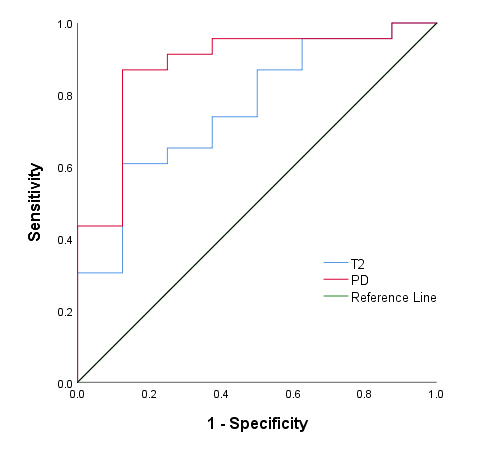
Figure 2 ROC cure analysis of the mean T2 and PD values for
differentiating low- from high-grade BCa. The area under the ROC curves for the mean T2 and PD values were 0.761,
and 0.880, respectively.