Lihua Chen1, Ailian Liu1, Pengyun Zhang1, Nila Mu1, Yunsong Liu1, Changjun Ma1, Jiazheng Wang2, Qingwei Song1, and Renwang Pu1
1The First Affiliated Hospital of Dalian Medical University, Dalian, China, 2Philips Healthcare, Beijing, China
1The First Affiliated Hospital of Dalian Medical University, Dalian, China, 2Philips Healthcare, Beijing, China
There were statistic differences in the APT and IVIM parameters between prostate cancer and prostatic hyperplasia. Higher diagnostic confidence was achieved using APT together with IVIM to differentiate between prostate cancer and prostatic hyperplasia.
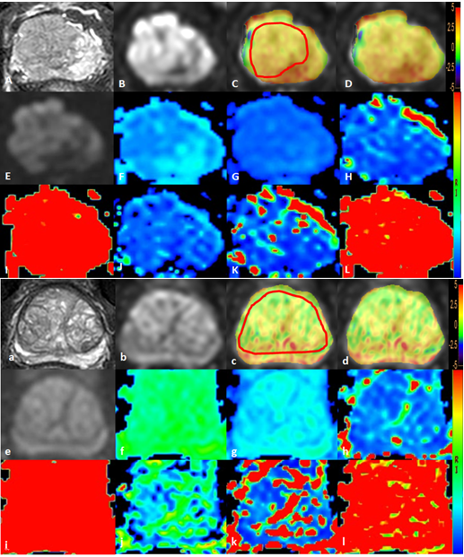
Figure 1 A 72-year-old man with PCa: T2WI image (A), DWI
image (B, the tumor was located in the right central zone), APTw image of the
prostate fused with DWI image (C, D, the placement of ROIs is as illustrated). The tumor showed the APTw value of 1.95 %. IVIM images as well as the derived maps
(E-L).
A 70-year-old man with BPH: T2WI image (a), DWI
image (b), APTw image of the prostate fused with DWI image (c, Dd, the placement of ROIs is as
illustrated). The prostate
showed the APTw value of 1.31 %. IVIM images as well as the derived maps (e-l).
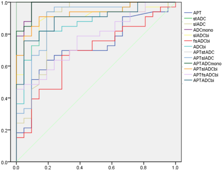
Figure 2 The ROC of all the parameters values in
differentiation of PCa and BPH.