Xiaobing Fan1, Aritrick Chatterjee1, Jay M Pittman1, Ambereen Yousuf1, Tatjana Antic2, Gregory S Karczmar1, and Aytekin Oto1
1Radiology, The University of Chicago, Chicago, IL, United States, 2Pathology, The University of Chicago, Chicago, IL, United States
1Radiology, The University of Chicago, Chicago, IL, United States, 2Pathology, The University of Chicago, Chicago, IL, United States
A split dose protocol for Dotarem
injection with ultrafast DCE-MRI sampling can improve detection of prostate
cancer by improving quantitative
analysis and providing sensitivity to water exchange.
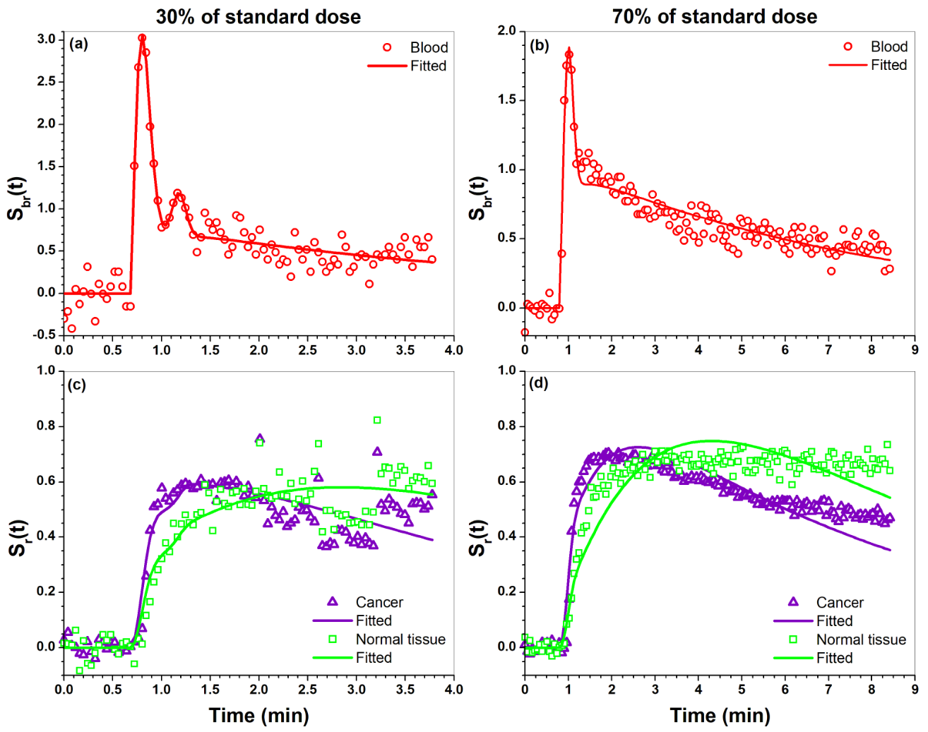
Figure 1. Plots of blood Sbr(t) (dots) obtained
from iliac artery and its EMM fits (line) for (a) 30% of standard dose and (b) 70%
dose of standard dose. (c) and (d) show plots of cancer and tissue Sr(t)
(dots) and their fits (lines) obtained from the SI-Tofts model for (c) 30% and
(d) 70% of standard dose.
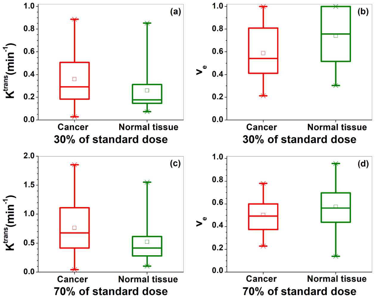
Figure 2. Box-plots of cancer (red) and
normal tissue (green) physiological parameters (a ,c) Ktrans and (b,
d) ve extracted from SI-Tofts model for the 30% (top row) and 70% (bottom
row) of standard dose DCE-MRI. The square
(□) indicates mean and the asterisks (*) indicate the upper and lower limits of
the data.