Anne Adlung1, Fabian Tollens2, Nadia Karina Paschke1, Jennifer Hümsch1, Niklas Westhoff3, Daniel Hausmann2,4, Lothar Rudi Schad1, Dominik Nörenberg2, and Frank Gerrit Zöllner1,5
1Computer Assisted Clinical Medicine, Medical Faculty Mannheim, Heidelberg University, Mannheim, Germany, 2Department of Radiology and Nuclear Medicine, Medical Faculty Mannheim, Heidelberg University, Mannheim, Germany, 3Department of Urology and Urosurgery, Medical Faculty Mannheim, Heidelberg University, Mannheim, Germany, 4Department of Radiology, Kantonspital Baden, Baden, Switzerland, 5Mannheim Institute for Intelligent System in Medicine, Medical Faculty Mannheim, Heidelberg University, Mannheim, Germany
1Computer Assisted Clinical Medicine, Medical Faculty Mannheim, Heidelberg University, Mannheim, Germany, 2Department of Radiology and Nuclear Medicine, Medical Faculty Mannheim, Heidelberg University, Mannheim, Germany, 3Department of Urology and Urosurgery, Medical Faculty Mannheim, Heidelberg University, Mannheim, Germany, 4Department of Radiology, Kantonspital Baden, Baden, Switzerland, 5Mannheim Institute for Intelligent System in Medicine, Medical Faculty Mannheim, Heidelberg University, Mannheim, Germany
This study investigates TSC quantification in the human prostate based
on internal references (iliac artery). 23Na-MRI of 19 male patients with suspected
PCa was included. No statistically significant differences were found compared
to external references.
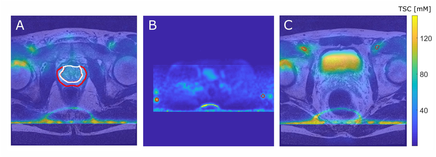
Figure 1:
A: T2w and coregistered 23Na MRI illustrated as overlay with the TZ (white) and the PZ (red).
B: TSC quantification based on reference vials prior to image co-registration (method 1, vials encirecled in red).
C: Quantification based on VOIs within the iliac artery after image co-registration to the T2w image (method 2, iliac arteries encircled in red).
For all three images a slice was chosen that best depicts the corresponding ROI. Images show high intensity lines at the bottom which are artifacts that were caused by the B1-corrections.
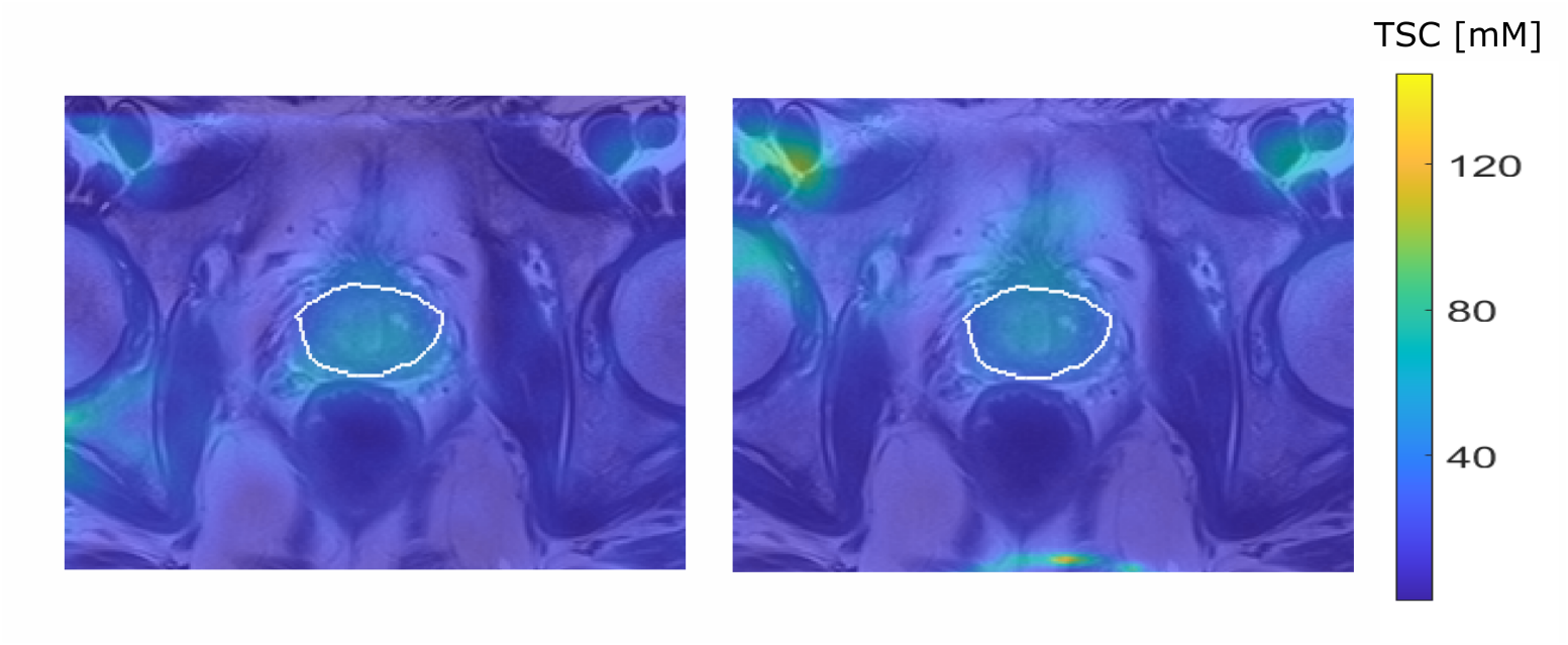
Figure 3: Quantified 23Na
MRI from one patient with a PI-RADs 5 lesion in the TZ, image focused on the prostate
region. TSC quantification based on method1 (left) and method2 (right).