Melline Gabrielle Maria Schilham1, Patrik Zamecnik1, Bas Israel1, Bastiaan Privé1, Mark Rijpkema1, Jelle Barentsz1, James Nagarajah1, Martin Gotthardt1, and Tom Scheenen1
1Medical Imaging, Radboudumc, Nijmegen, Netherlands
1Medical Imaging, Radboudumc, Nijmegen, Netherlands
Nano-MRI identifies significantly
more small suspicious lymph nodes compared to PSMA-
PET/CT in the same patient.
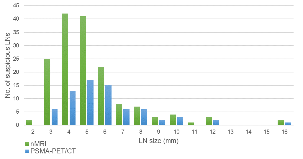
Size distribution of suspicious lymph nodes as
detected by nanoparticle-enhanced MRI (green) and prostate-specific membrane
antigen PET/CT (blue).
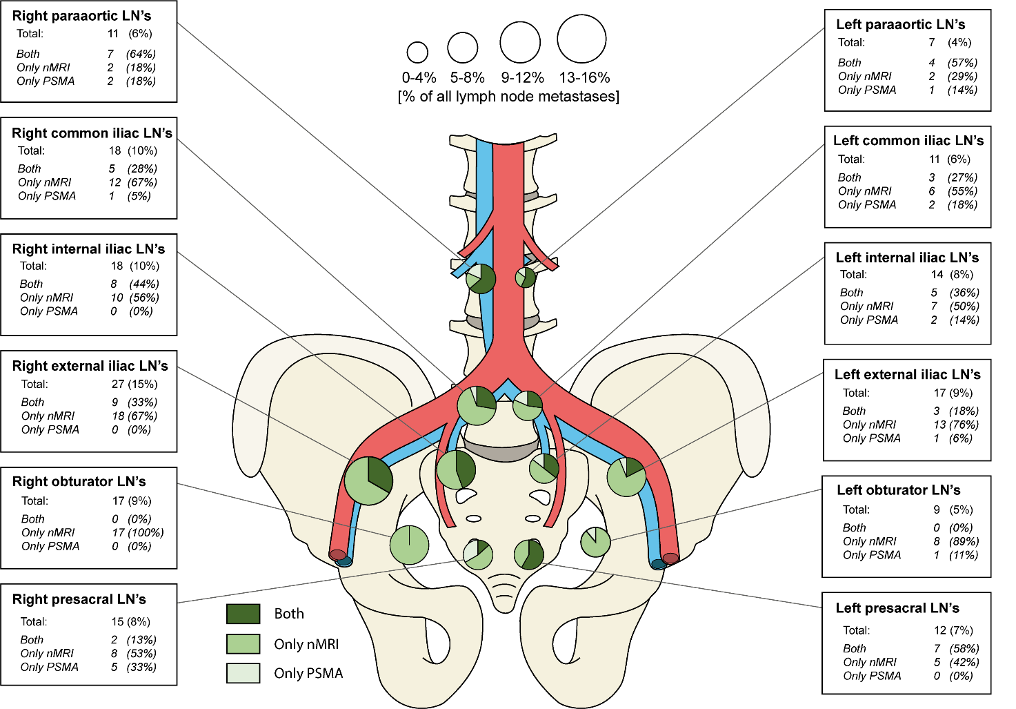
FIGURE 3. Anatomic distribution of identified suspicious lymph nodes as
detected by nanoparticle-enhanced MRI and prostate-specific membrane antigen
PET/CT.