Xiaoping Zhu1, Daniel Lewis2, Ka-Loh Li1, Sha Zhao3, Timothy Cootes1, Andrew King2, David Coope2, and Alan Jackson3
1DIIDS, University of Manchester, Manchester, United Kingdom, 2Neurosurgery, Salford Royal NHS Foundation Trust, Manchester, United Kingdom, 3University of Manchester, Manchester, United Kingdom
1DIIDS, University of Manchester, Manchester, United Kingdom, 2Neurosurgery, Salford Royal NHS Foundation Trust, Manchester, United Kingdom, 3University of Manchester, Manchester, United Kingdom
In brain DCE-MRI the superior sagittal sinus can provide a superior global VIF compared to large intracranial arteries, demonstrating lower noise, higher bolus peak-amplitude and greater sensitivity to inter-individual changes in plasma contrast-agent concentration.
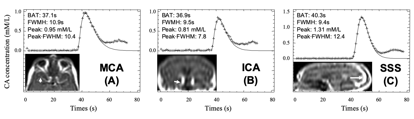
Figure 2: Typical plasma contrast-agent concentration curves Cp(t) extracted from the horizontal segment of the middle cerebral artery (arrow head), MCA (A); vertical segment of the ICA (short arrow), carotid syphon (B); and vertical segment of the SSS (long arrow) (C) following a bolus injection of 0.023 mmol/kg of GBCA. The 1st-pass data are fitted using a gamma variate function, which excludes contrast-agent bolus recirculation. 3D T1-weighted gradient recalled echo mages obtained from a patient with a sporadic VS scanned at 1.5T.
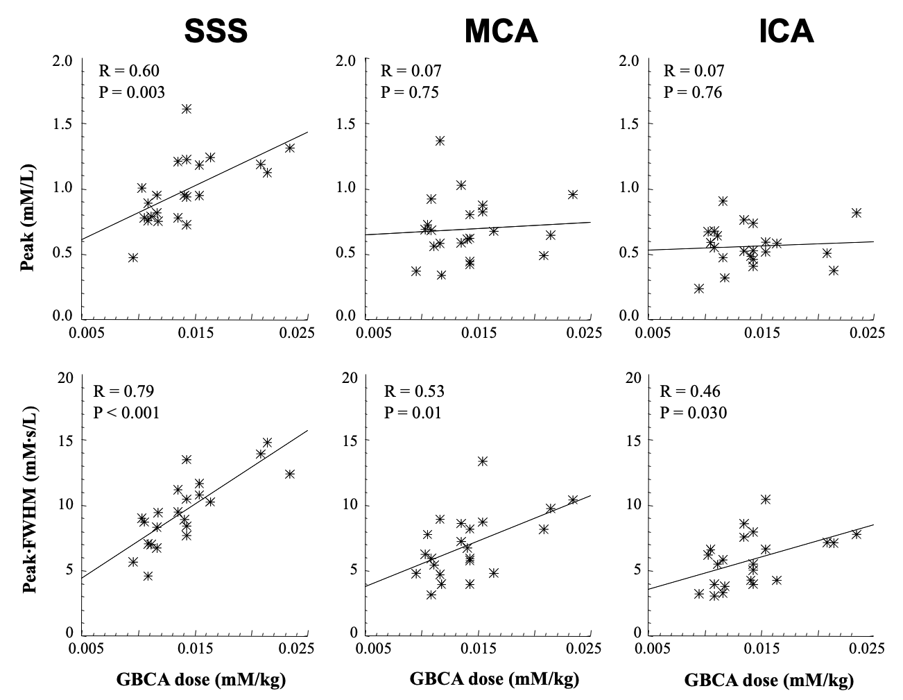
Figure 3: Relationship between extracted VIF features and administered GBCA dose for VIFSSS, VIFMCA and VIFICA. Top row: Scatter plots of VIF peak (mM/L) vs administered GBCA dose (mM/kg). Bottom row: Scatter plots of area (= peak∙FWHM: full-width at half-maximum, mM·s /L) of VIF vs administered GBCA dose.