Qing Fu1,2, Qi-guang Cheng1,2, Xiao-yong Zhang3, Xiang-chuang Kong1,2, Ding-xi Liu1,2, Yi-hao Guo4, John Grinstead5, Zi-qiao Lei1,2, and Chuan-sheng Zheng1,2
1Department of Radiology, Union Hospital, Tongji Medical College, Huazhong University of Science and Technology, Wuhan, China, 2Hubei Province Key Laboratory of Molecular Imaging, Wuhan, China, 3Centers for Biomedical Engineering, University of Science and Technology of China, Hefei, China, 4MR Collaboration, Siemens Healthcare Ltd., Guangzhou, China, 5Siemens Medical Solutions USA, Inc., Portland, OR, United States
1Department of Radiology, Union Hospital, Tongji Medical College, Huazhong University of Science and Technology, Wuhan, China, 2Hubei Province Key Laboratory of Molecular Imaging, Wuhan, China, 3Centers for Biomedical Engineering, University of Science and Technology of China, Hefei, China, 4MR Collaboration, Siemens Healthcare Ltd., Guangzhou, China, 5Siemens Medical Solutions USA, Inc., Portland, OR, United States
Small cerebral metastases can be difficult to differentiate from blood vessels and surrounding parenchyma. Here we show that DANTE-SPACE outperforms both conventional MPRAGE and an advanced technique (PETRA) through blood vessel suppression.
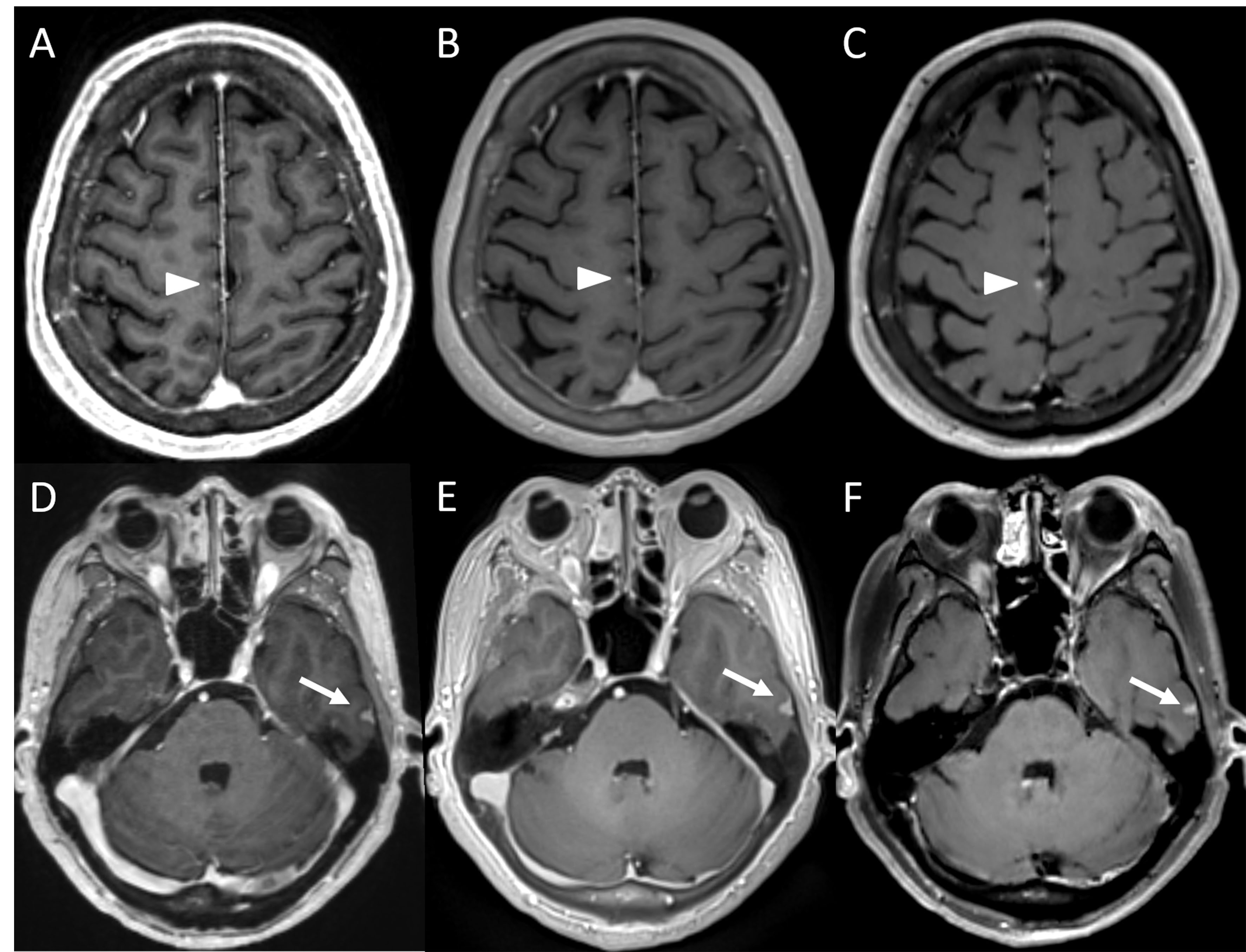
Figure 1. Representative images of MPRAGE (A, D), PETRA (B, E) and DANTE-SPACE (C, F) of a 57-year-old male with cerebral metastases of lung cancer. A-C: a well-enhanced lesion (arrowheads) near the cerebral falx in the right frontal lobe is seen in all sequences. The lesion is more conspicuous with DANTE-SPACE due to blood vessel suppression and high CNR, and could have been mislabeled as a small vessel with MPRAGE and PETRA. D-F: a small homogeneously meningioma in the left temporal lobe was displayed clearly in all sequences with clear margin
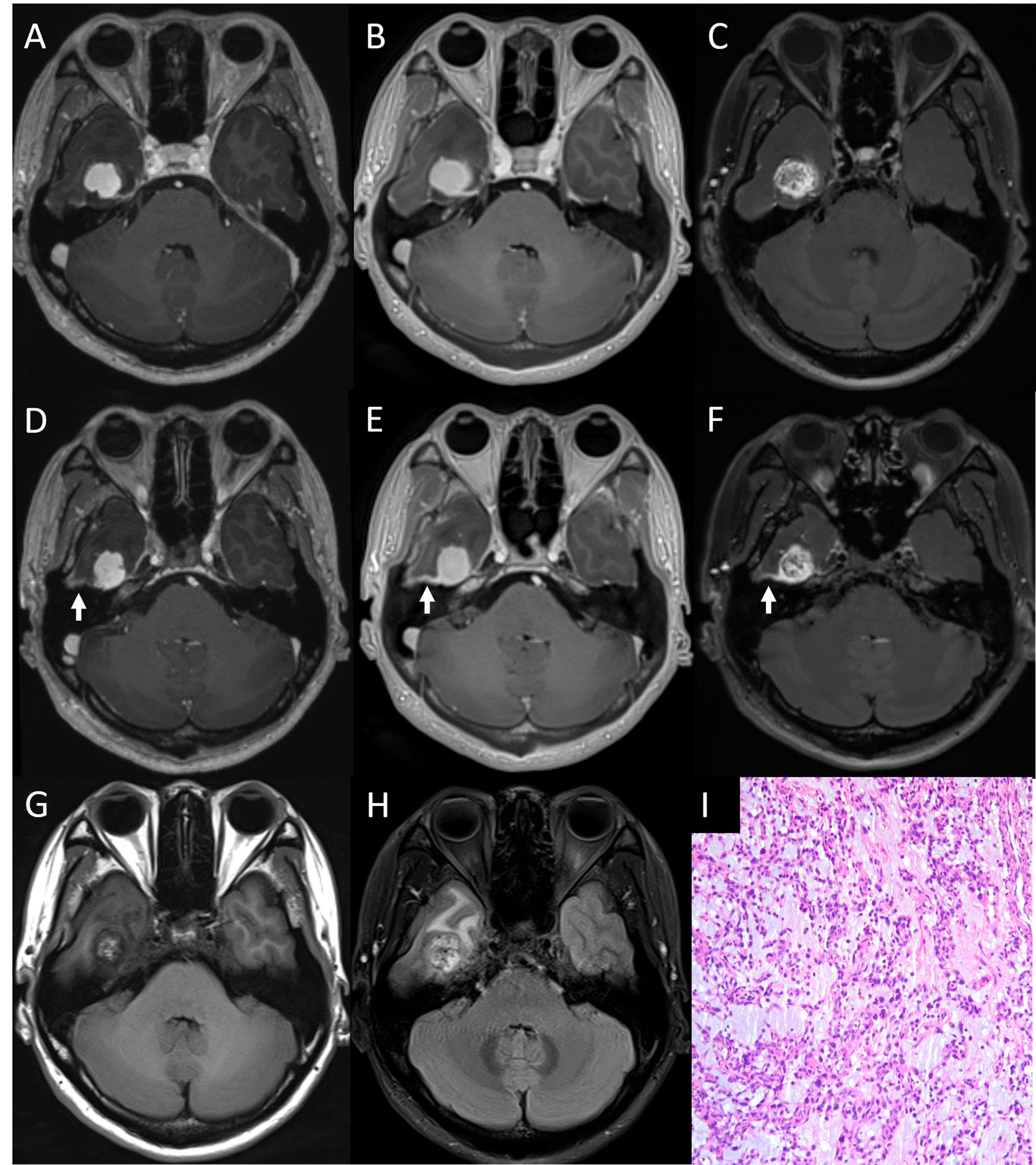
Figure 2. Post-contrast T1-weighted images of an enhancing tumor in right temporal lobe in a 37-year-old female. The signal intensity within this tumor was relatively homogeneous in MPRAGE (A) and PETRA (B), but it was heterogeneous in DANTE-SPACE (C). The enhancing effect (arrows) of adjacent meninges is more continuous in PETRA. The tumor signal from DANTE-SPACE (C) closely resembles pre-contrast T1WI and T2WI (G, H), and it was identified as a myopericytoma by surgical pathology (I).