Augusto Lio M. Goncalves Filho1,2, John Conklin1,2, Chanon Ngamsombat3, Stephen F. Cauley2, Wei Liu4, Daniel N. Splitthoff5, Wei-Ching Lo6, John E. Kirsch1, Pamela W. Schaefer1, Otto Rapalino1, and Susie Y. Huang1,2
1Department of Radiology, Massachusetts General Hospital, Boston, MA, United States, 2Department of Radiology, Athinoula A. Martinos Center for Biomedical Imaging, Charlestown, MA, United States, 3Department of Radiology, Siriraj Hospital, Bangkok, Thailand, 4Siemens Shenzhen Magnetic Resonance Ltd., Shenzhen, China, 5Siemens Healthcare GmbH, Erlangen, Germany, 6Siemens Medical Solutions Inc., Boston, MA, United States
1Department of Radiology, Massachusetts General Hospital, Boston, MA, United States, 2Department of Radiology, Athinoula A. Martinos Center for Biomedical Imaging, Charlestown, MA, United States, 3Department of Radiology, Siriraj Hospital, Bangkok, Thailand, 4Siemens Shenzhen Magnetic Resonance Ltd., Shenzhen, China, 5Siemens Healthcare GmbH, Erlangen, Germany, 6Siemens Medical Solutions Inc., Boston, MA, United States
An accelerated post-contrast Wave-CAIPI 3D T1 MPRAGE sequence is
non-inferior to the standard T1 MPRAGE for detection of intracranial enhancing lesions
in 3T MRI while providing a 2-fold reduction in acquisition time and being less
sensitive to motion artifacts.
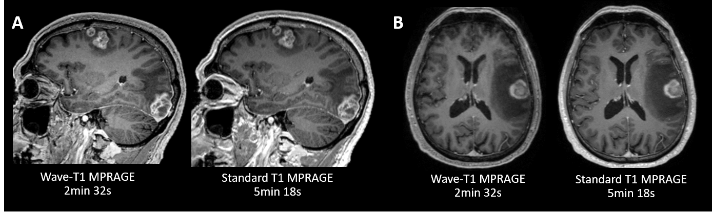
Figure 2. Representative
images showing a comparison of post-contrast Wave-CAIPI T1 MPRAGE and standard
T1 MPRAGE sequences. (A) A 56-year-old male with brain metastases from
squamous-cell carcinoma. (B) A 71-year-old male with brain metastases
from non-small cell lung cancer.
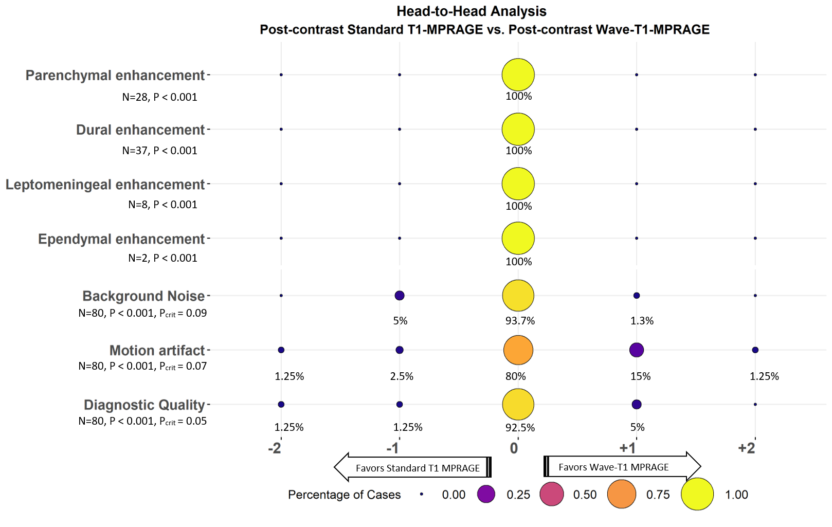
Figure 1. Balloon plot showing the results of the
head-to-head comparison of post-contrast standard T1 MPRAGE and post-contrast
Wave-CAIPI T1 MPRAGE for visualization of abnormal intracranial enhancing
lesions, the perception of noise, presence of artifacts due to motion, and the
overall diagnostic quality. A zero-score indicates equivalency, negative scores
(left) favor standard T1 MPRAGE, and positive scores (right) favor Wave-T1
MPRAGE.