Zahra Shams1, Sarah M. Jacobs1, Jan W. Dankbaar1, Changho Choi2, Dennis W.J. Klomp1, Jannie P. Wijnen1, and Evita C. Wiegers1
1Department of Radiology, University Medical Center Utrecht, Utrecht, Netherlands, 2Advanced Imaging Research Center, University of Texas Southwestern Medical Center, Dallas, TX, United States
1Department of Radiology, University Medical Center Utrecht, Utrecht, Netherlands, 2Advanced Imaging Research Center, University of Texas Southwestern Medical Center, Dallas, TX, United States
We implemented 2HG MR spectroscopy in the
clinical routine of the glioma patients. We showed technical feasibility, the
success rate and robustness of the protocol.
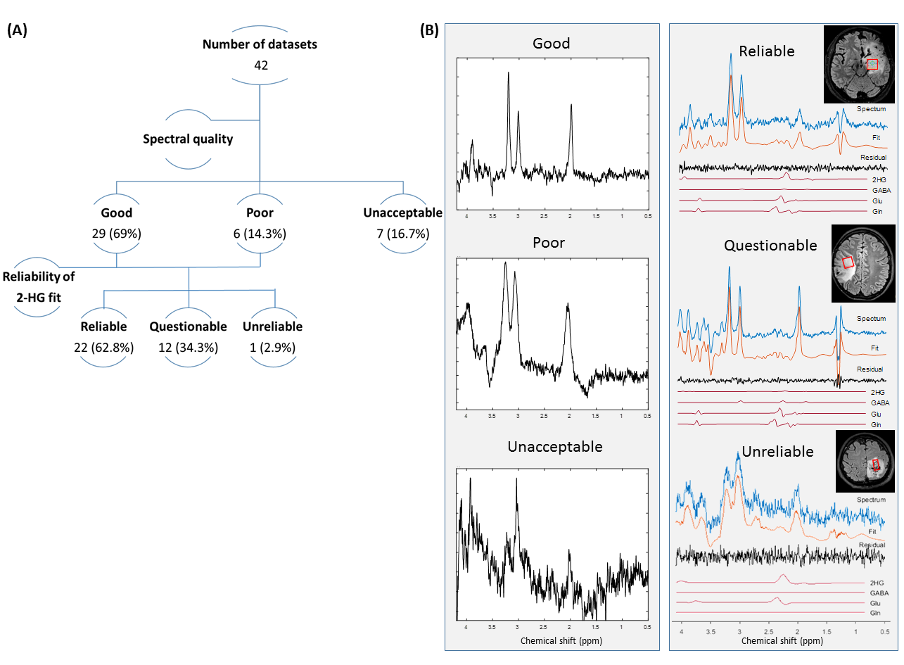
Figure 1. Evaluation of the spectra based on quality and
reliability of the 2HG fit. Examples of each group have been shown in (B).
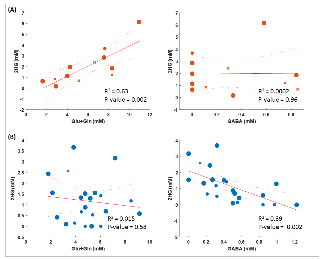
Figure 3. The relationship between 2HG concentration and the
concentration of Glu+Gln and GABA in ‘Questionable’ (A) and ‘Reliable’ (B)
fits. ×: information on voxel location in the tumor tissue is not available.
Small marker size: no tumor (likelihood of 0-25%); big marker size: tumor
(likelihood of 50-100%).