Chan Hong Moon1, Krystof Bankiewicz2, Paul Larson2, Adrian Kells3, Alastair J. Martin2, Stephen Mancuso4, and Mark Richardson5
1University of Pittsburgh, Pittsburgh, PA, United States, 2University of California San Francisco, San Francisco, CA, United States, 3Voyager Therapeutics Inc., Cambridge, MA, United States, 4UPMC, Pittsburgh, PA, United States, 5Massachusetts General Hospital, Boston, MA, United States
1University of Pittsburgh, Pittsburgh, PA, United States, 2University of California San Francisco, San Francisco, CA, United States, 3Voyager Therapeutics Inc., Cambridge, MA, United States, 4UPMC, Pittsburgh, PA, United States, 5Massachusetts General Hospital, Boston, MA, United States
In this study, we developed new dual-contrast MPRAGE and maximized T1 contrast in putamen as well as good anatomy of whole brain without additional acquisition compared to conventional MPRAGE. The proposed methods were applied to 1.5T iMRI during neurosurgical procedure of PD patients.
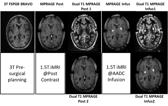
Fig. 3 T1 anatomy MRI; (from left) Pre-surgical @3T, Post contrast at1.5T (MPRAGE, dual T1 MPRAGEs) and AADC infusion at1.5T (MPRAGE, dual T1 MPRAGEs). Note that new dual T1 MPRAGE at1.5T can provide higher contrast of Put than that at 3T. In addition, dual T1 MPRAGE provides good whole brain anatomy. White spots are the targeted infusion regions.
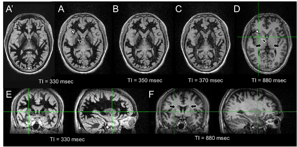
Fig. 2 3D MPRAFGE MR images at effective TI 330 (A and A’), 350 (B), 370 (C) and 880 msec (D) at kz = 0; only axial images are shown. The images in A and A’ were acquired at a week apart sessions. Black arrows indicate the aliasing artifact in conventional MPRAGE image. Note that the signal in the white mater at optimal TI ~330 msec is well suppressed and the contrast of putamen (white arrowhead) and glubus pallidus (black arrowhead) vs. white mater is much higher than those of conventional MPRAGE image at effective TI 880 msec. In addition, the optimal TI MPRAGE MRI is reproducible.