Chuanke Hou1, Guanzhong Gong1, Weiqiang Dou2, and Yong Yin1
1Department of Radiation Physics, Shandong Cancer Hospital and Institute, Shandong First Medical University and Shandong Academy of Medical Sciences, Jinan, China, 2GE Healthcare, MR Research China, Beijing, China
1Department of Radiation Physics, Shandong Cancer Hospital and Institute, Shandong First Medical University and Shandong Academy of Medical Sciences, Jinan, China, 2GE Healthcare, MR Research China, Beijing, China
3D-Araterial Spine Labeling can be used to segment high and low cerebral blood flow sub-volumes. Then the plan with maximum dose constraint performed better for dose escalation in low cerebral blood flow (hypoxic) sub-volumes without increasing the dose delivered to organs at risk.
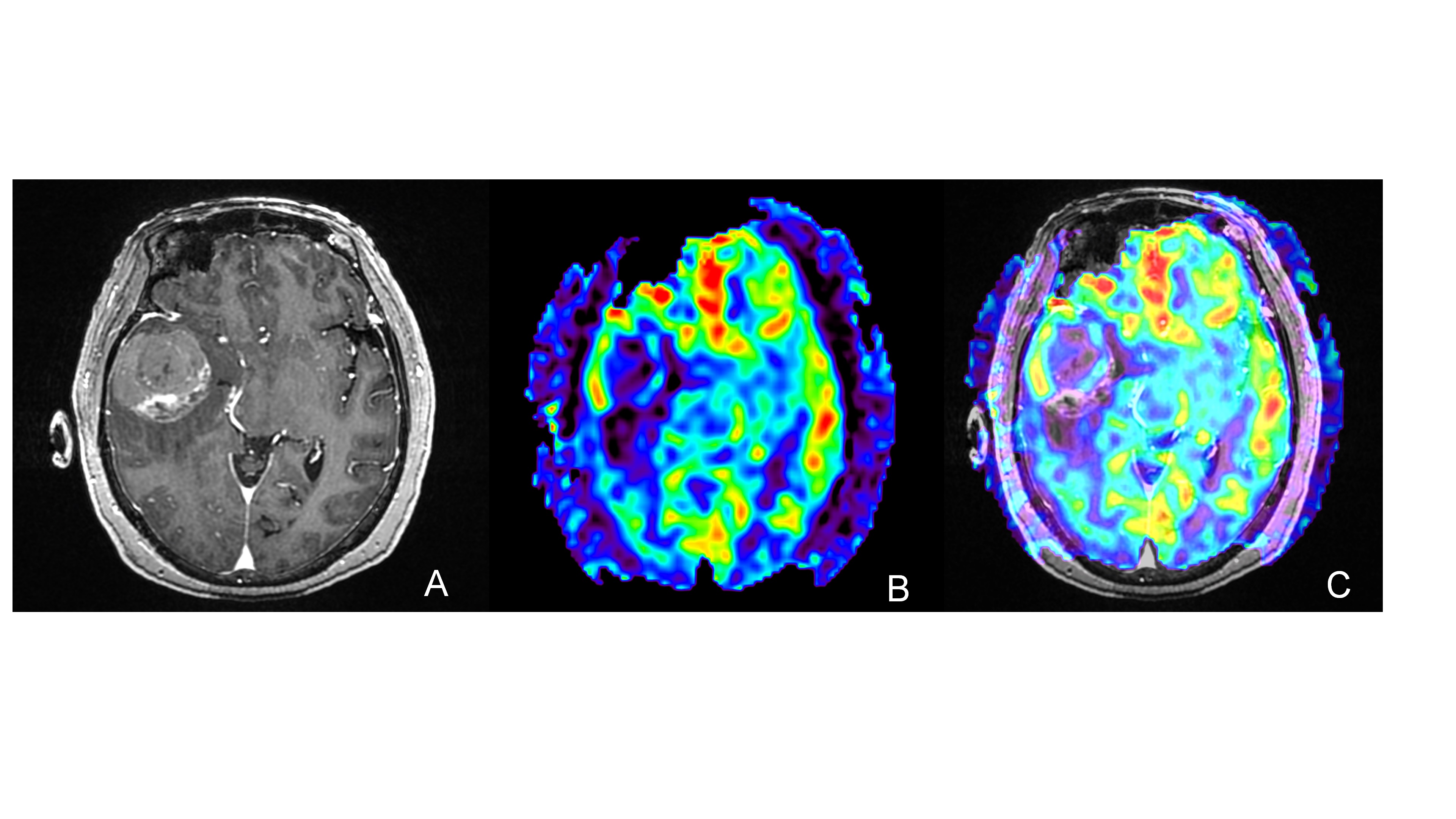
Figure1 Different tumor information was shown between contrast-enhanced T1W images and 3D-ASL images. A: contrast-enhanced T1W images; B:3D-ASL; C: 3D-ASL fusion registration to contrast-enhanced T1W images.According to the enhanced area shown in the figure A, figure B showed the uneven distribution of CBF in tumor. The high cerebral blood flow area was mainly located on the right side of the enhanced edge, while the low cerebral blood flow area and the enhanced area overlap in a large region. Moreover, the fusion image C showed this result more clearly.
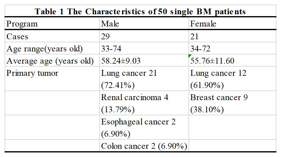
Table 1 The Characteristics of 50 single BM patients