Timothy Bray1,2, Alan Bainbridge1,3, Susan Jawad2, Sofia Otero2, Timothy J Beale2, Sumandeep Kaur2, Mark McGurk4, Margaret A Hall-Craggs1,2, and Simon Morley2
1Centre for Medical Imaging, University College London, London, United Kingdom, 2Department of Imaging, University College London Hospital, London, United Kingdom, 3Medical Physics, University College London Hospital, London, United Kingdom, 4Head and Neck Academic Centre, University College London, London, United Kingdom
1Centre for Medical Imaging, University College London, London, United Kingdom, 2Department of Imaging, University College London Hospital, London, United Kingdom, 3Medical Physics, University College London Hospital, London, United Kingdom, 4Head and Neck Academic Centre, University College London, London, United Kingdom
An approach to 'neurographic' imaging of the extracranial facial nerve at high resolution using variable flip angle turbo spin echo imaging is proposed. This method depicts the nerve as a low-signal structure (‘black nerve’) against the high-signal parotid parenchyma (‘white parotid’).
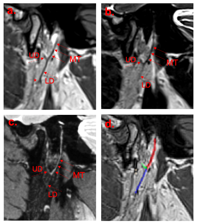
Figure 2: Example images. Methods 1, 2 and 3 are shown in (a), (b) and (c) respectively. An example of a segmentation mask is shown in (d): the main trunk is segmented in red, the upper division in green and the lower division in blue.
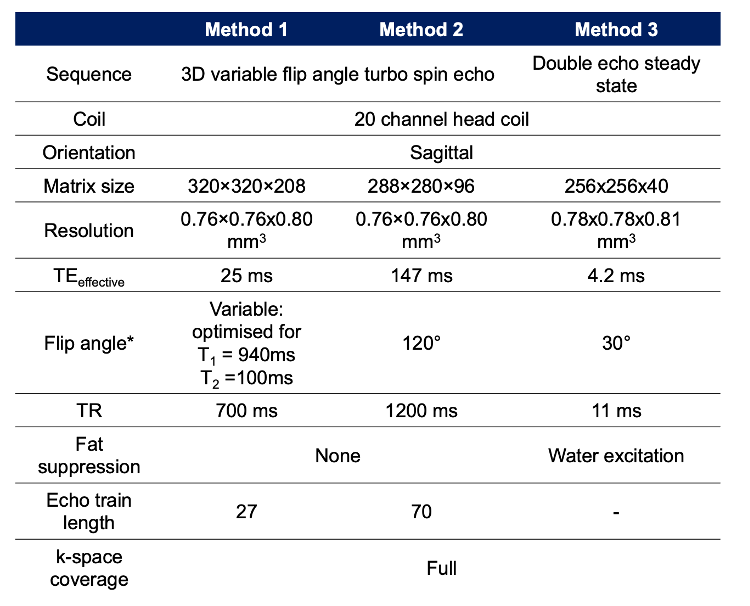
Figure 1: MRI sequences and acquisition parameters.