David Volders1, James Rioux1, Steven Beyea1, Chris V Bowen2, and Elena Adela Cora1
1Diagnostic Radiology, Nova Scotia Health, Halifax, NS, Canada, 2Diagnostic Imaging, Nova Scotia Health, Halifax, NS, Canada
1Diagnostic Radiology, Nova Scotia Health, Halifax, NS, Canada, 2Diagnostic Imaging, Nova Scotia Health, Halifax, NS, Canada
Evaluation of temporal bone MRI scans using a 0.5T high performance gradient head only system reveals high quality visualization of clinically relevant structures using 3D bSSFP imaging.
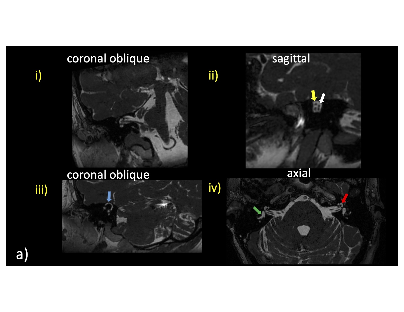
Figure 1: Representative cross-sectional 3D bSSFP images for a patient at 0.5T (a) and 3T (b) reformatted into various labeled planes (i.-iv.). Key anatomical structures being rated by 2 board certified radiologists are shown. Blue arrow: superior semicircular canal; Yellow arrow: facial (top) and cochear (bottom) nerve; White arrow: superior vestibular (top) and inferior vestibular (bottom) nerve; Green arrow: vestibule; Red arrow: cochlea.
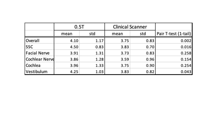
Table 1: Statistical comparison of 0.5T vs clinical field strength scans