Dadi Zhao1,2, James T. Grist1,2, Heather E.L. Rose1,2, Huijun Li2, Lesley MacPherson2, Yu Sun1,2, and Andrew C. Peet1,2
1Institute of Cancer and Genomic Sciences, University of Birmingham, Birmingham, United Kingdom, 2Department of Oncology, Birmingham Children's Hospital, Birmingham, United Kingdom
1Institute of Cancer and Genomic Sciences, University of Birmingham, Birmingham, United Kingdom, 2Department of Oncology, Birmingham Children's Hospital, Birmingham, United Kingdom
Multi-modal functional imaging provides improved classification performance for childhood brain tumours. The combination of diffusion weighted imaging and noise-suppressed proton magnetic resonance spectroscopy shows promising for classifying imbalance childhood brain tumours.
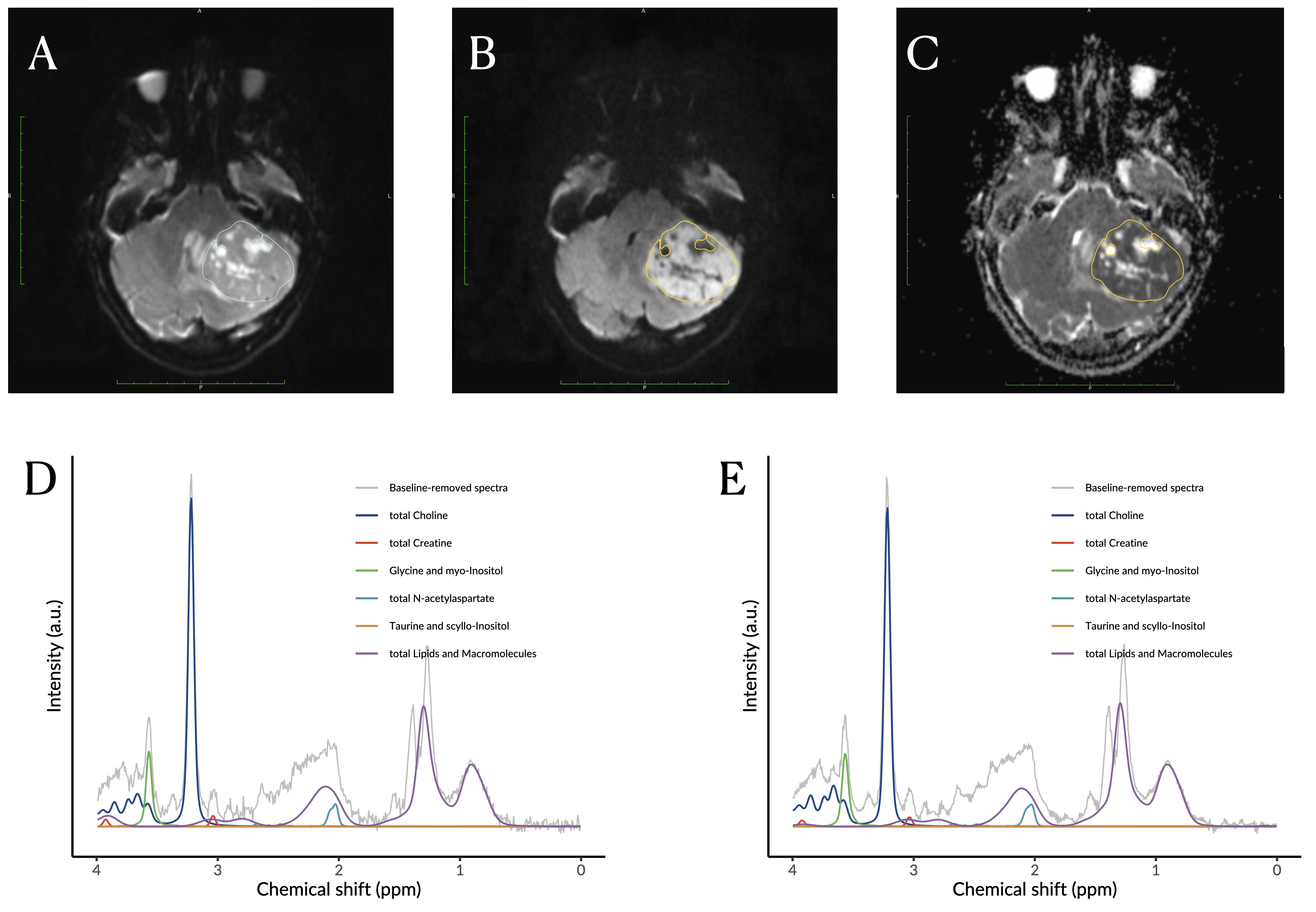
Images showing the acquired b0 (A), b1000 (B) and apparent diffusion coefficient (C) images with the region of interest for brain tumour as well as the noisy (D) and noise-suppressed (E) proton magnetic resonance spectroscopy of a patient with a medulloblastoma.
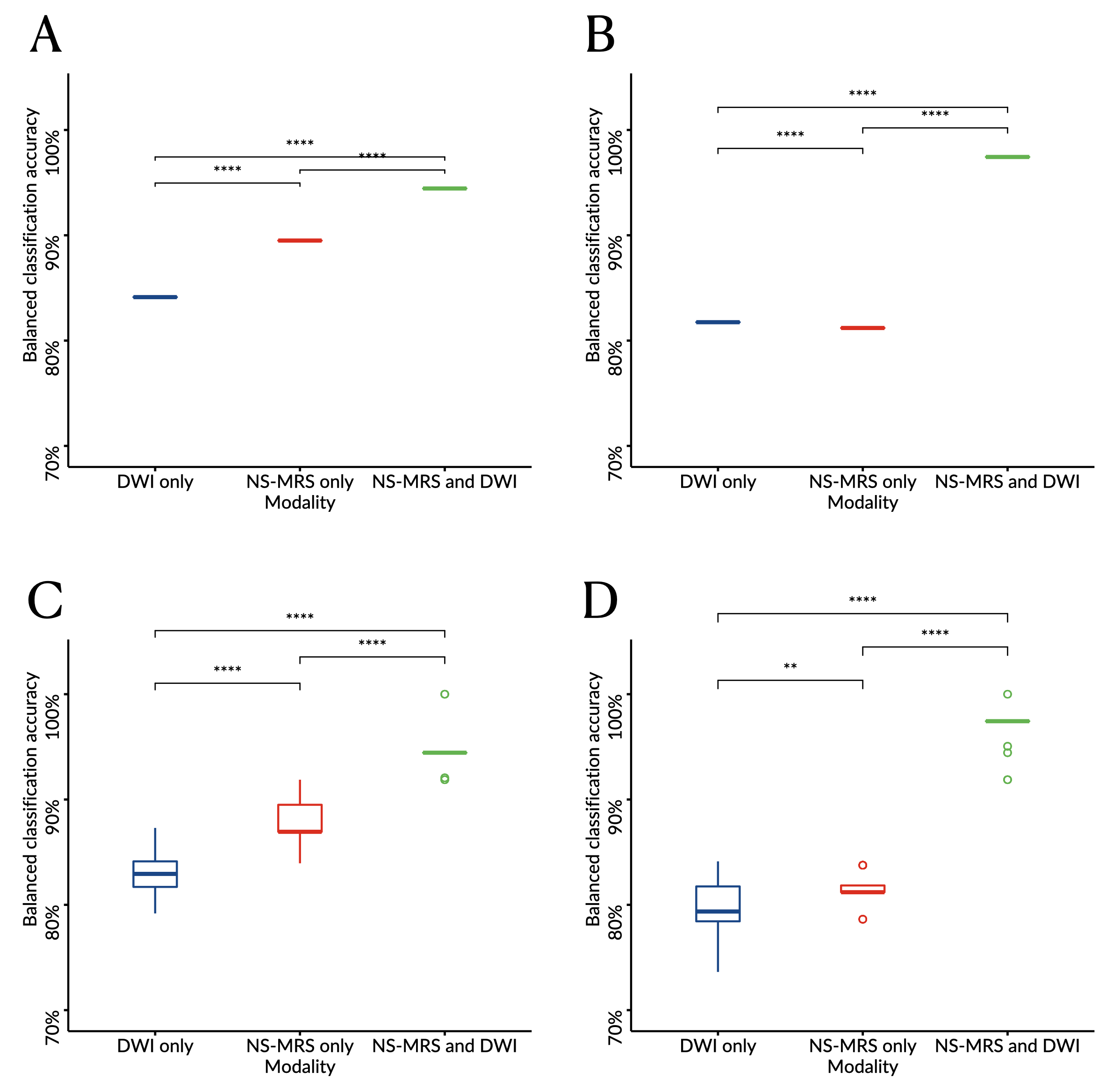
Boxplots comparing the balanced classification accuracy estimated through leave-one-out (A-B) or six-fold (C-D) cross validation and determined through linear discriminant analysis (A, C) or support vector machine (B, D) from diffusion weighted imaging (DWI) only, noise-suppressed proton magnetic resonance spectroscopy (NS-MRS) only, or combining NS-MRS and DWI.
Levels of significance: *, P<.05; **, P<.01; ***, P<.001; ****, P<.0001.