Yoshimi Anzai1, John A Roberts2, Seong-Eun Kim2, Ying Hitchcock3, Richard Wiggins1, Nousheen Alasti2, and Eugene Kholmovski2
1Department of Radiology and Imaging Sciences, University of Utah, Salt Lake City, UT, United States, 2UCAIR, Department of Radiology and Imaging Sciences, University of Utah, Salt Lake City, UT, United States, 3Radiation oncology, University of Utah, Salt Lake City, UT, United States
1Department of Radiology and Imaging Sciences, University of Utah, Salt Lake City, UT, United States, 2UCAIR, Department of Radiology and Imaging Sciences, University of Utah, Salt Lake City, UT, United States, 3Radiation oncology, University of Utah, Salt Lake City, UT, United States
A composite color map combining quantitative information from multiparametric imaging (oxygen-enhanced BOLD, DWI, and FDG-PET) has the potential to reveal the spatial distribution of the area of hypoxia within a tumor.
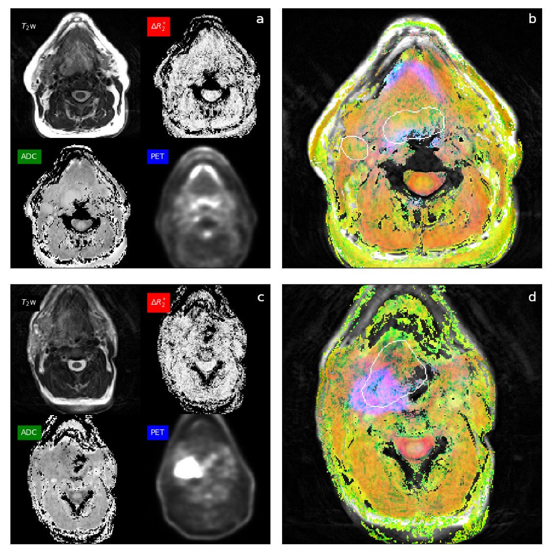
Fig 3. The color map of the patient (a, b) shows a green color of the base of tongue tumor and right lymph node metastases suggestive of low ADC value with non-zero ΔR2*, oxygenated tumor. The color map of another patient (c, d) of right tonsillar carcinoma shows pink-magenta color posteriorly, suggestive of near-zero ΔR2* and high PET activity, features of hypoxic tumor. This patient had early recurrence and disease-related mortality
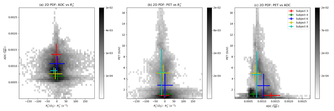
Fig 2. 2D scatter histograms comparing (a) ADC to DR2*, (b) FDG-PET to ΔR2*, and (c) FDG-PET to ADC. A large standard deviation (length of vertical and horizontal bars) and ΔR2* are observed in a patient with recurrence (subject 6).