Qing Fu1,2, Xiang-chuang Kong1,2, Ding-xi Liu1,2, Yi-hao Guo3, Kun Zhou4, Zi-qiao Lei1,2, and Chuan-sheng Zheng1,2
1Department of Radiology, Union Hospital, Tongji Medical College, Huazhong University of Science and Technology, Wuhan, China, 2Hubei Province Key Laboratory of Molecular Imaging, Wuhan 430022, China, Wuhan, China, 3MR Collaboration, Siemens Healthcare Ltd., Guangzhou, China., Guangzhou, China, 4Siemens Shenzhen Magnetic Resonance Ltd., Shenzhen, China., Shenzhen, China
1Department of Radiology, Union Hospital, Tongji Medical College, Huazhong University of Science and Technology, Wuhan, China, 2Hubei Province Key Laboratory of Molecular Imaging, Wuhan 430022, China, Wuhan, China, 3MR Collaboration, Siemens Healthcare Ltd., Guangzhou, China., Guangzhou, China, 4Siemens Shenzhen Magnetic Resonance Ltd., Shenzhen, China., Shenzhen, China
2D turbo gradient and spin echo BLADE-DWI was superior to readout-segmented echo-planar-DWI in depicting orbital tumors with reduced susceptibility artifacts.
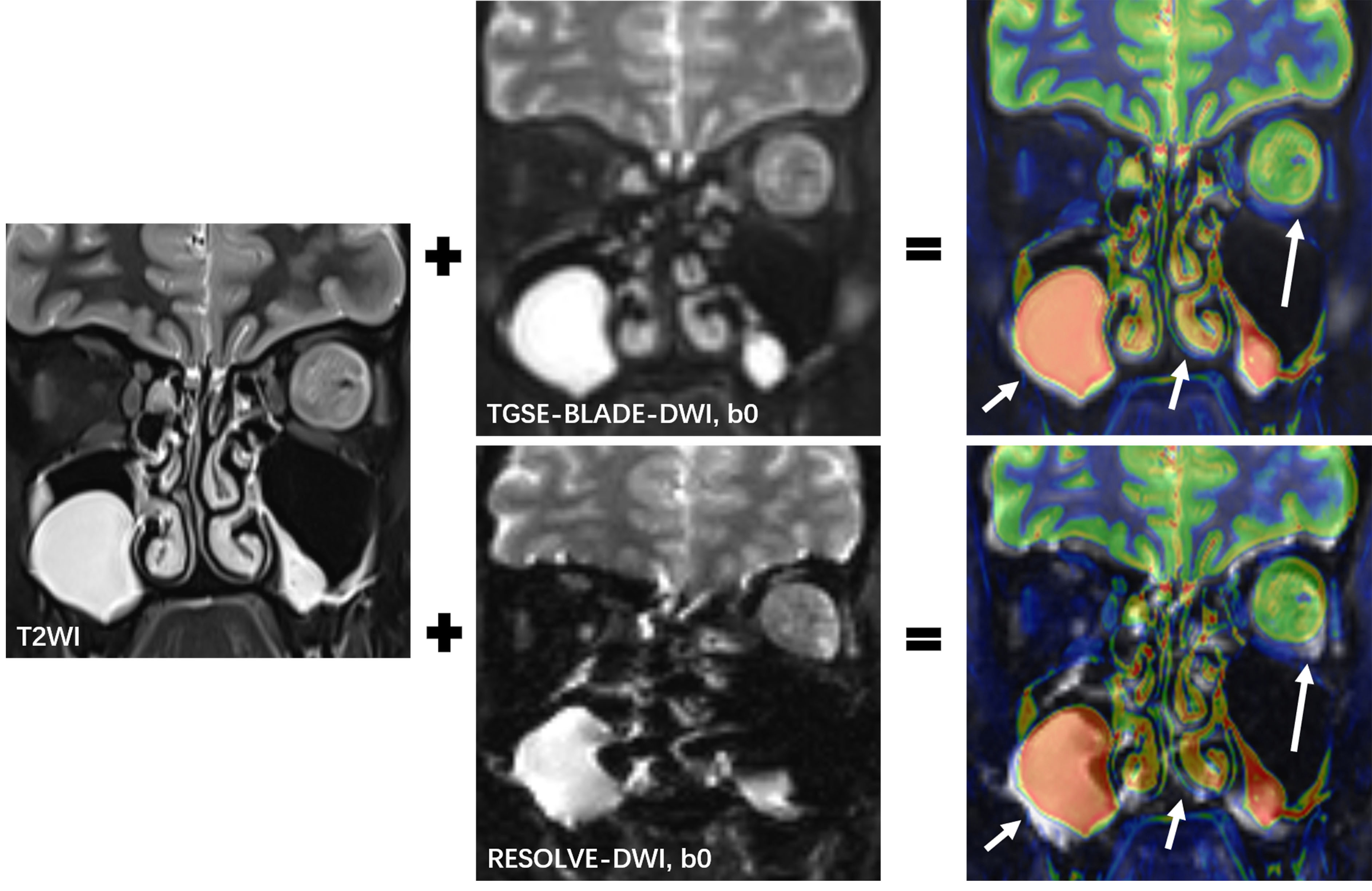
Figure 1. T2-weighted
imaging (T2WI), b0 images of 2D TGSE-BLADE-DWI, and RESOLVE-DWI were fused. Color-
and gray-coated images were derived from the T2WI and the two DWI b0 images,
respectively. The T2WI and TGSE-BLADE-DWI b0 images matched well, showing the left
orbital tumor (long arrows), ethmoid and maxillary sinuses (short arrows). The T2WI
and RESOLVE-DWI b0 images were mismatched due to geometric distortions.
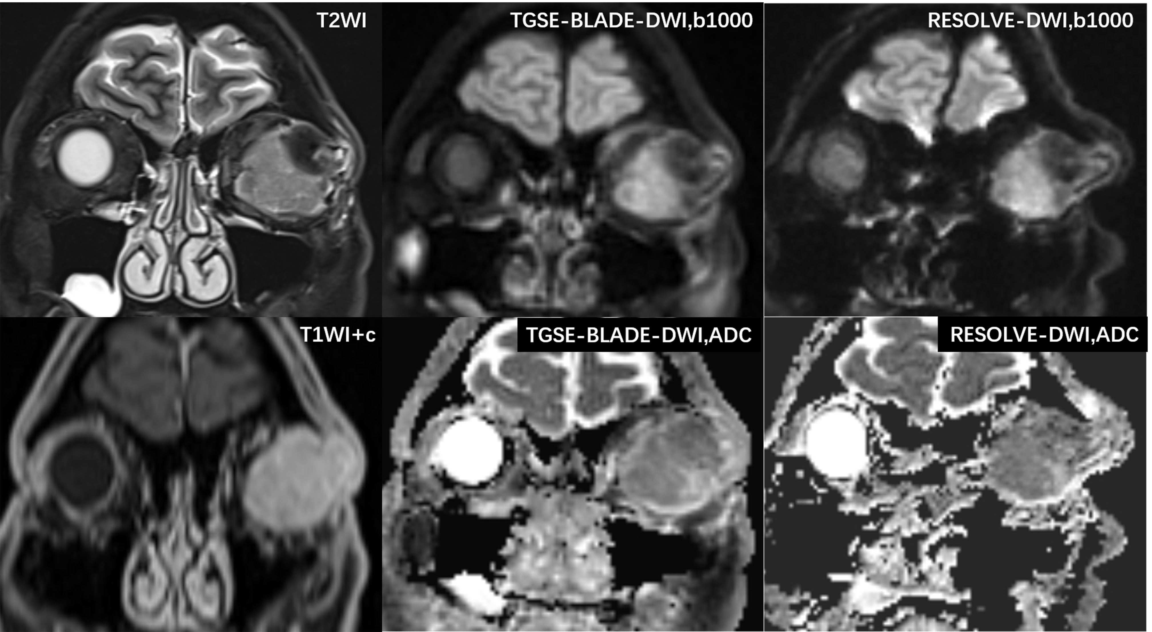
Figure 2. A 50-year-old male with a left orbital tumor. Compared with the T2-weighted imaging (T2WI) images and contrast-enhanced T1WI images, this lesion could be visualized clearly with TGSE-BLADE-DWI; no geometric distortions were seen. Slight distortions were seen with RESOLVE-DWI. More geometric distortions and ghosting artifacts were observed in the frontal lobe and ethmoid sinus with RESOLVE-DWI.