Qing Fu1,2, Xiang-chuang Kong1,2, Ding-xi Liu1,2, Yi-hao Guo3, Kun Zhou4, Zi-qiao Lei1,2, and Chuan-sheng Zheng1,2
1Department of Radiology, Union Hospital, Tongji Medical College, Huazhong University of Science and Technology, Wuhan, China, 2Hubei Province Key Laboratory of Molecular Imaging, Wuhan 430022, China, Wuhan, China, 3MR Collaboration, Siemens Healthcare Ltd., Guangzhou, China., Guangzhou, China, 4Siemens Shenzhen Magnetic Resonance Ltd., Shenzhen, China., Shenzhen, China
1Department of Radiology, Union Hospital, Tongji Medical College, Huazhong University of Science and Technology, Wuhan, China, 2Hubei Province Key Laboratory of Molecular Imaging, Wuhan 430022, China, Wuhan, China, 3MR Collaboration, Siemens Healthcare Ltd., Guangzhou, China., Guangzhou, China, 4Siemens Shenzhen Magnetic Resonance Ltd., Shenzhen, China., Shenzhen, China
2D turbo gradient- and spin-echo-BLADE diffusion-weighted imaging (DWI) was found to be superior to readout-segmented echo-planar-DWI and single-shot echo-planar-DWI in depicting and diagnosing cerebellopontine angle tumors.
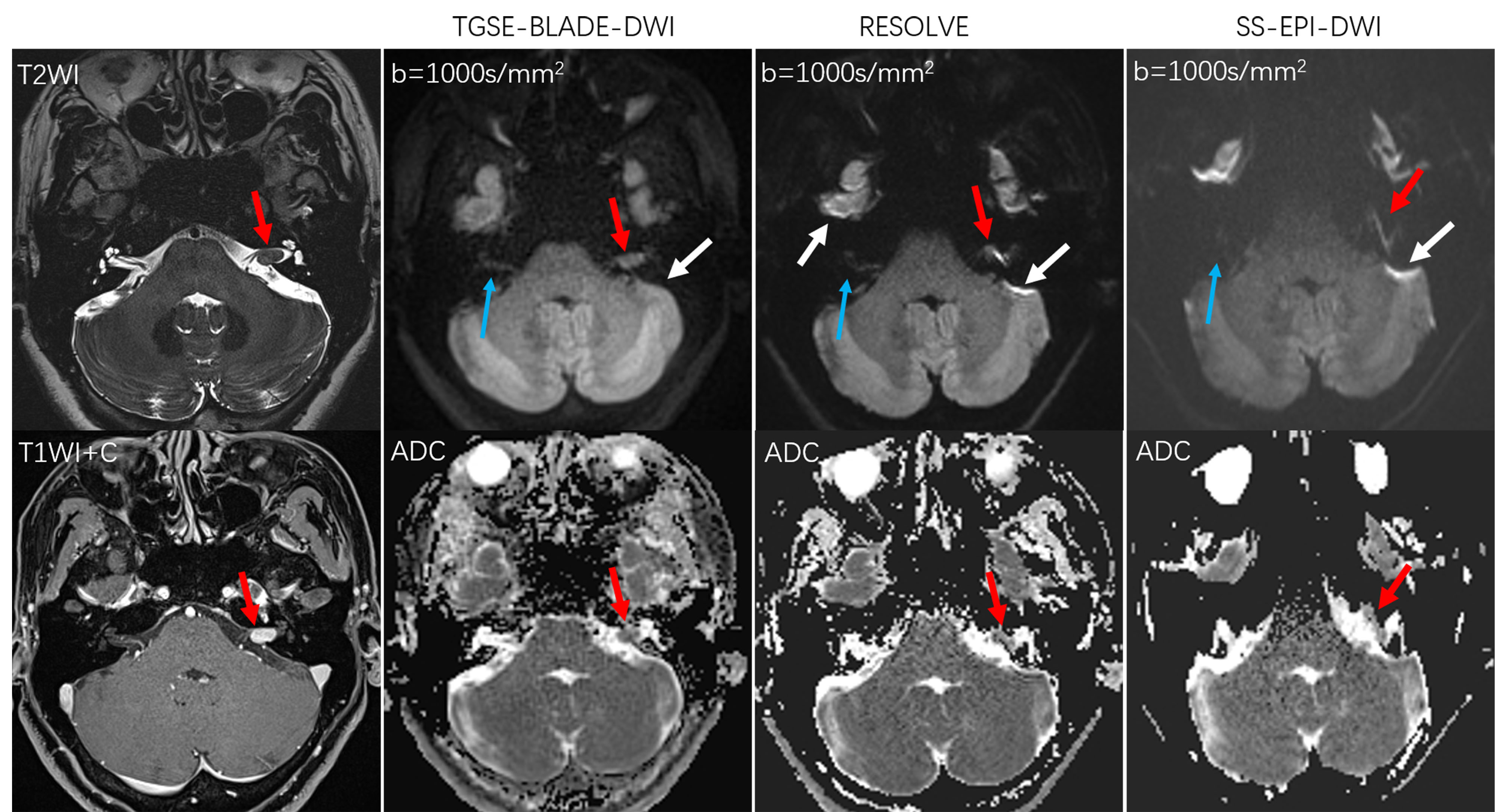
Figure 1. A 50-year-old female with a left internal auditory canal acoustic neuroma (red arrows). The lesion is clearly visible on the TGSE-BLADE-DWI scans without geometric distortion, but with moderate and severe distortions on the RESOLVE-DWI and SS-EPI-DWI scans, respectively, interfering with lesion detection. Ghosting artifacts (white arrows) were absent at bone-air interfaces and anatomic structural identifications (blue arrows) were superior on the TGSE-BLADE-DWI scans compared with the other two DWI methods.
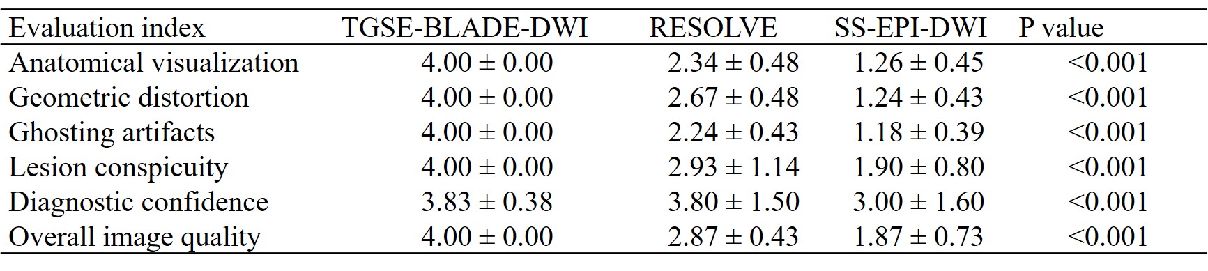
Table 3. A comparison of the subjective evaluation results among the three diffusion-weighted imaging methods.