Yutaka Hamatani1, Kayoko Abe2, Masami Yoneyama3, Jaladhar Neelavalli4, Yasuhiro Goto1, Isao Shiina1, Kazuo Kodaira1, Takumi Ogawa1, Mamoru Takeyama1, Isao Tanaka1, and Shuji Sakai2
1Department of Radioligical Services, Tokyo Women's Medical University Hospital, Tokyo, Japan, 2Department of Diagnostic imaging & Nuclear Medicine, Tokyo Women's Medical University Hospital, Tokyo, Japan, 3Philips Japan, Tokyo, Japan, 4Philips Healthcare, Bangalore, India
1Department of Radioligical Services, Tokyo Women's Medical University Hospital, Tokyo, Japan, 2Department of Diagnostic imaging & Nuclear Medicine, Tokyo Women's Medical University Hospital, Tokyo, Japan, 3Philips Japan, Tokyo, Japan, 4Philips Healthcare, Bangalore, India
IRIS (Image
Reconstruction using Image-space Sampling Function) is one of multi shot echo
planar EPI-DWIs combined with phase correction, and it was the best sequence to
visualize the optic nerve, compared other conventional DWI techniques.
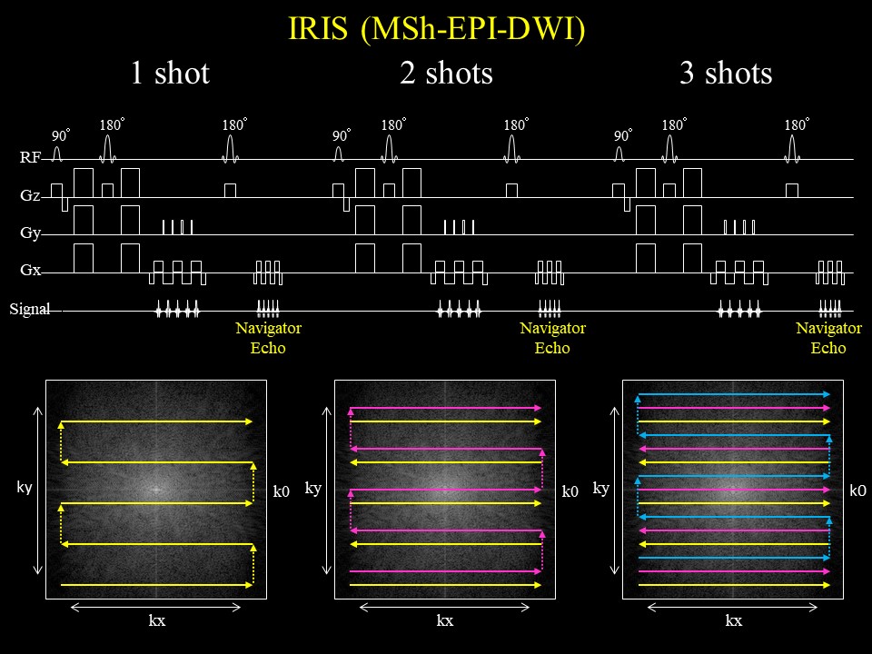
Figure 1.The IRIS
sequence chart. IRIS adopts multi shot acquisition which signal collection into
k-space is divided in the phase direction. Multi shot acquisition tends to
cause ghost artifacts, so IRIS applies navigator-echo for each shot to make phase
corrections.
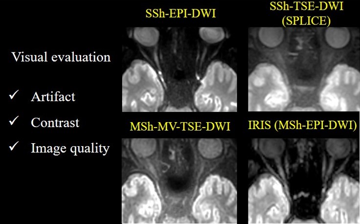
Figure 2. DWI of the optic
nerve on SSh EPI-DWI, SSh TSE-DWI, MSh. TSE-DWI, and IRIS. IRIS shows the least
artifacts and image distortion, and the optic nerve was demonstrated with the
highest image contrast on IRIS.