1Radiology, Fujita Health University School of Medicine, Toyoake, Japan, 2Canon Medical System Corporation, Otawara, Japan, 3Joint Research Laboratory of Advanced Medical Imaging, Fujita Health University School of Medicine, Toyoake, Japan, 4Fujita Health University Hospital, Toyoake, Japan
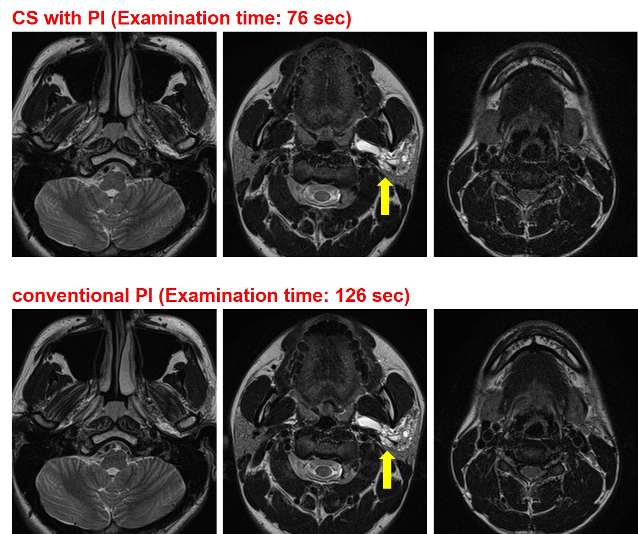
Figure1. 21-year-old male with lymphatic malformation (L to R: cranial to caudal level).
T2-weighted images obtained with CS with PI and with conventional PI clearly demonstrate the multiple cystic lesions extending from parotid space to parapharyngeal space (arrows). Neither image showed significant artifacts and both had the same overall image quality. The examination time of CS with PI (76 sec) was much shorter than that of conventional PI (126 sec).
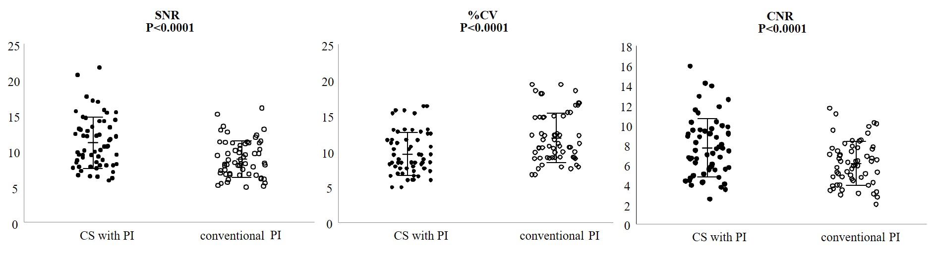
Figure 2. Results of comparison for each quantitative image quality index among CS with PI and conventional PI.
SNR of CS with PI (11.2±3.6, mean ± standard deviation) was significantly higher than that of conventional PI (8.9±2.6, p<0.0001). %CV of CS with PI (9.6±3.0) was significantly lower than that of conventional PI (11.9±3.5, p<0.0001). CNR of CS with PI (7.7±2.9) was significantly higher than that of conventional PI (6.1±2.2, p<0.0001).