Timothy Bray1,2, Ruaridh Gollifer1,3, Simon Morley2, Susan Jawad2, Roganie Govender4, Stuart A Taylor1,2, Nicholas Hamilton4, and David Atkinson1
1Centre for Medical Imaging, University College London, London, United Kingdom, 2Department of Imaging, University College London Hospital, London, United Kingdom, 3Medical Physics, University College London Hospital, London, United Kingdom, 4Head and Neck Academic Centre, University College London, London, United Kingdom
1Centre for Medical Imaging, University College London, London, United Kingdom, 2Department of Imaging, University College London Hospital, London, United Kingdom, 3Medical Physics, University College London Hospital, London, United Kingdom, 4Head and Neck Academic Centre, University College London, London, United Kingdom
We describe and evalute a method for imaging and analysis of pharyngeal and laryngeal motion during the swallow, based on joint image registration and modelling of intensity changes over a dynamic image series acquired at high temporal resolution.
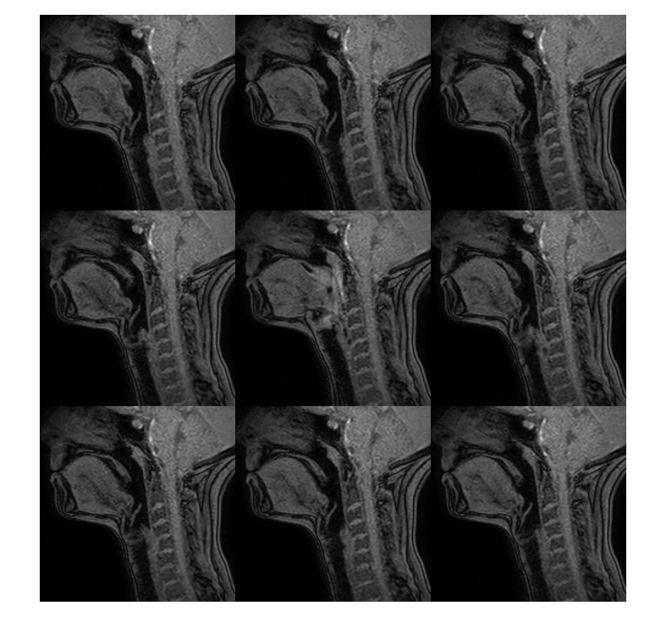
Figure 2 – Example time series of swallow. Selected images from a healthy volunteer (every fifth slice) are shown from left to right, starting with the top row. In the middle slices, the pharynx constricts and the hyoid and pre-epiglottic fat elevate.
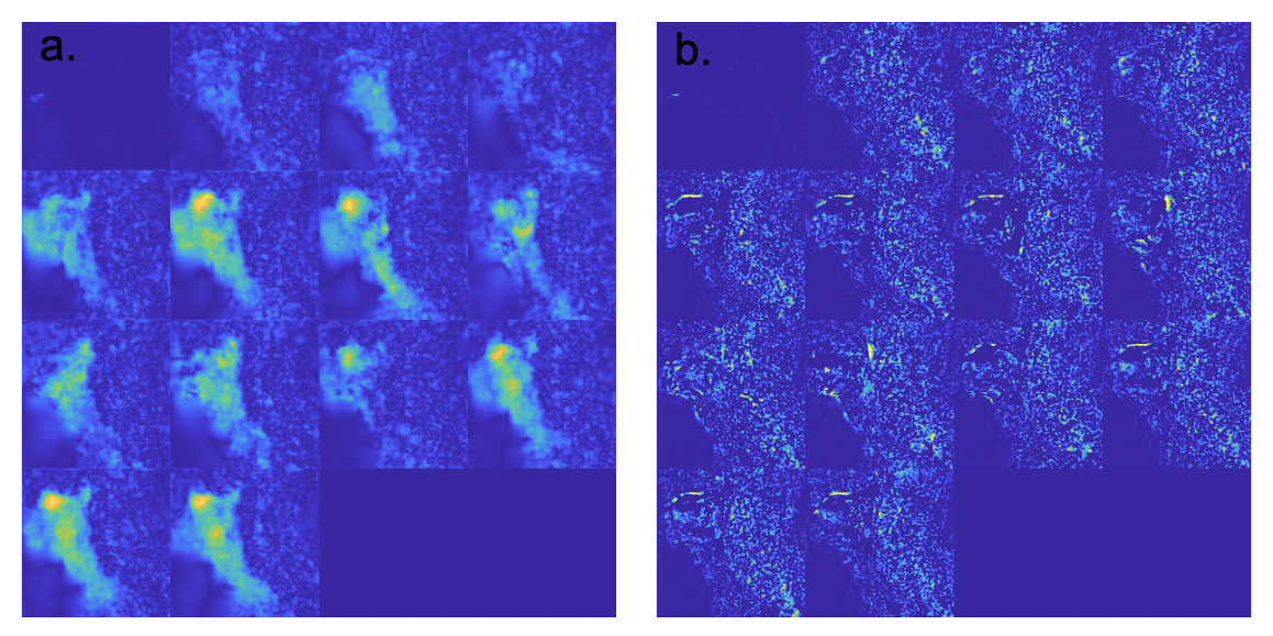
Figure 3 – Deformation maps and intensity change maps. Deformation magnitude is shown in (a) and is largest in the region of the tongue base and larynx. Intensity changes are shown in (b); in this case there are small intensity changes in the pharynx due to unsaturated tissue being pulled into the imaging slice.