Kilian Stumpf1, Mariam Seyfang2, Bernd Georg Lapatki2, and Volker Rasche1
1Department of Internal Medicine II, Ulm University Medical Center, Ulm, Germany, 2Department of Orthodontics, Ulm University Medical Center, Ulm, Germany
1Department of Internal Medicine II, Ulm University Medical Center, Ulm, Germany, 2Department of Orthodontics, Ulm University Medical Center, Ulm, Germany
A real-time radial
tiny golden angle sequence is applied for imaging of the temporomandibular
joint. Its use for visualizing anterior disc displacements
and assisting in diagnosis and treatment planning and evaluation is
demonstrated.
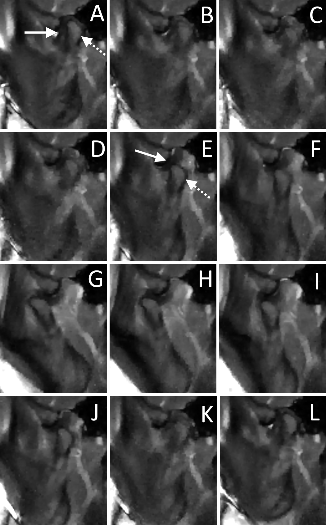
Figure 2: When starting the movement from a protrusive mandibular position, the sliding window
reconstruction with a frame rate of 125 ms allowed the visualization of the repositioning
of the condyle (dotted arrow) on the articular disc (solid arrow) (B-E) with
subsequent full opening of the mandible (G). During the closing motion the
condyle can once again be observed to slip down from the articular disc (J-L).
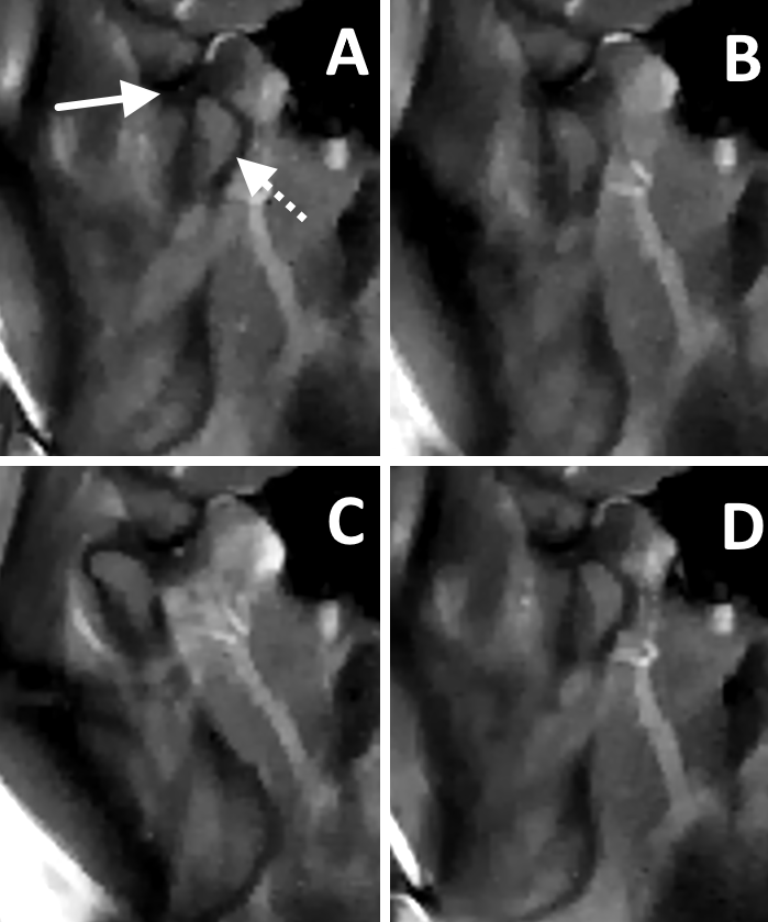
Figure 3: Visualization of the jaw motion with an incorporated occlusal splint. Starting in a therapeutic, more anterior and caudal
initial position with the condyle (dotted arrow) already positioned on the
articular disc (solid arrow), no additional repositioning or disc displacement was
observed during the entire motion of the jaw.