Jun Ma1,2, Bernhard Gruber3,4, Xinqiang Yan5, and William Grissom2
1Department of Radiology, Stanford University, Stanford, CA, United States, 2Department of Biomedical Engineering, Vanderbilt University, Nashville, TN, United States, 3A. A. Martinos Center for Biomedical Imaging, Massachusetts General Hospital, Harvard Medical School, Charlestown, MA, United States, 4Division MR Physics, Center for Medical Physics and Biomedical Engineering, Medical University Vienna, Vienna, Austria, 5Department of Radiology and Radiological Sciences, Vanderbilt University, Nashville, TN, United States
1Department of Radiology, Stanford University, Stanford, CA, United States, 2Department of Biomedical Engineering, Vanderbilt University, Nashville, TN, United States, 3A. A. Martinos Center for Biomedical Imaging, Massachusetts General Hospital, Harvard Medical School, Charlestown, MA, United States, 4Division MR Physics, Center for Medical Physics and Biomedical Engineering, Medical University Vienna, Vienna, Austria, 5Department of Radiology and Radiological Sciences, Vanderbilt University, Nashville, TN, United States
A k-space domain pTx pulse design algorithm is proposed,
which reduces the computation time of an example 3D pTx pulse design problem by
~80%, compared to a conventional spatial domain method.
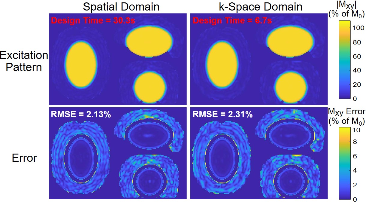
Fig 2: Normalized
excitation patterns (top row) and error maps (bottom row) in central slices for k-space domain (left) and spatial domain
designs (right). The calculated RMSEs were 2.13% (spatial) and 2.31%
(k-space), indicating uniform inner volume excitation
while maintaining the outer volume intact. The parallelized k-space domain
design required 6.7 s computation versus 30.3 s for the spatial
domain method, a 78% decrease. The size of the system matrix (W) of the
k-space domain design was 0.8 Gb, while the system matrix of the spatial domain
design had a size of 79 Gb, a 99% decrease.
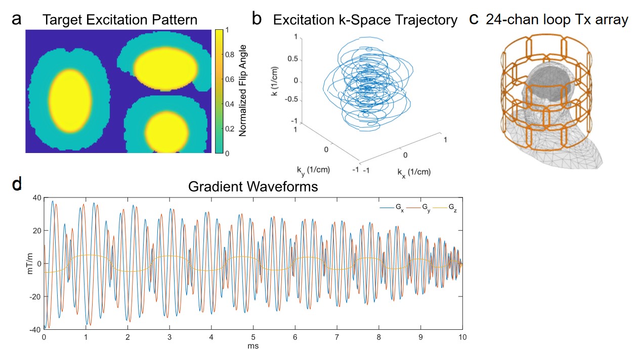
Fig 1: (a) Central slices of
the target excitation pattern used for all pulse designs, which comprised
an ellipse centered on the ventricles with AP/HF/LR semi-axes of 4.8/3.2/3.2
cm. (b) The SPINS trajectory used in the designs. (c)
The 24-channel loop Tx array that was simulated in a human head model to obtain B1+ maps. The array has diameter 32 cm, height 28 cm, and 16×11 cm rectangular loops in 3 rows of 8. (d) 10 ms
minimum time gradient waveforms that produce the SPINS trajectory, subject to
the scanner's gradient amplitude and slew rate constraints of 200 mT/m and 700
T/m/s, respectively.