Ziwei Zhao1, Nam G. Lee2, and Krishna S. Nayak1,2
1Ming Hsieh Department of Electrical and Computer Engineering, Viterbi School of Engineering, University of Southern California, Los Angeles, CA, United States, 2Department of Biomedical Engineering, Viterbi School of Engineering, University of Southern California, Los Angeles, CA, United States
1Ming Hsieh Department of Electrical and Computer Engineering, Viterbi School of Engineering, University of Southern California, Los Angeles, CA, United States, 2Department of Biomedical Engineering, Viterbi School of Engineering, University of Southern California, Los Angeles, CA, United States
Simulations indicate that velocity-selective RF pulses for intracranial MRA achieve comparable performance at 0.55T compared to 3T. We anticipate 50%-60% reduced signal loss and/or 22%-38% sharper velocity selection, potentially improving the detection of slow flow and distal vessels.
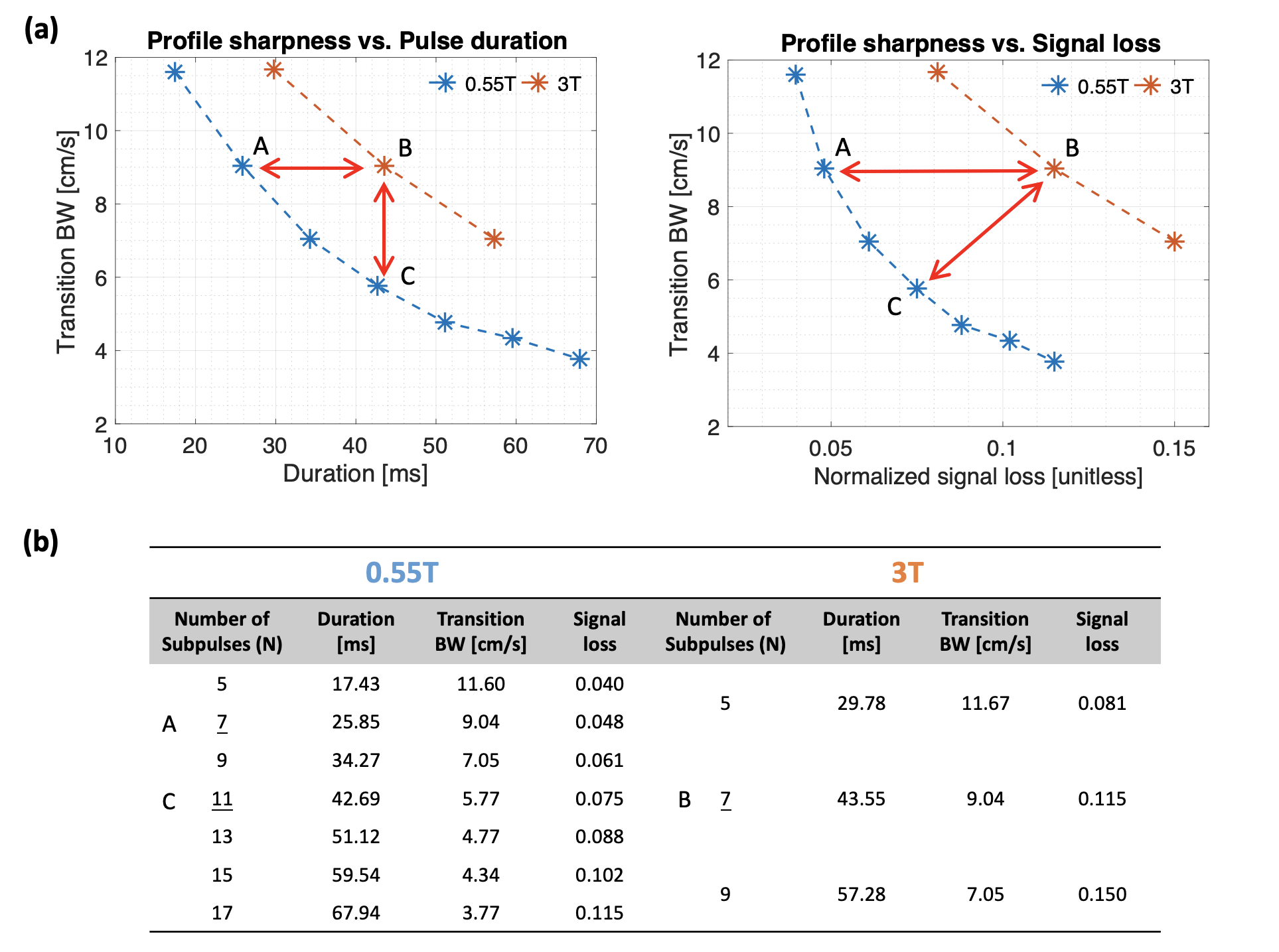
Figure 2. Pulse Performance Tradeoff. (a) Velocity profile sharpness vs. pulse duration (left) and signal loss (right) with (b) detailed parameters of plotted designs. T1 and T2 values were used as 1121ms, 263ms at 0.55T; and 1650ms, 150ms at 3T, respectively. Normalized signal loss is defined as (M0-Mz)/M0. Note that 0.55T system (blue) shows higher fidelities of tradeoffs. The RF and gradient waveforms of two representative designs (marked “A” and “B” asterisks) are plotted in Figure 1.
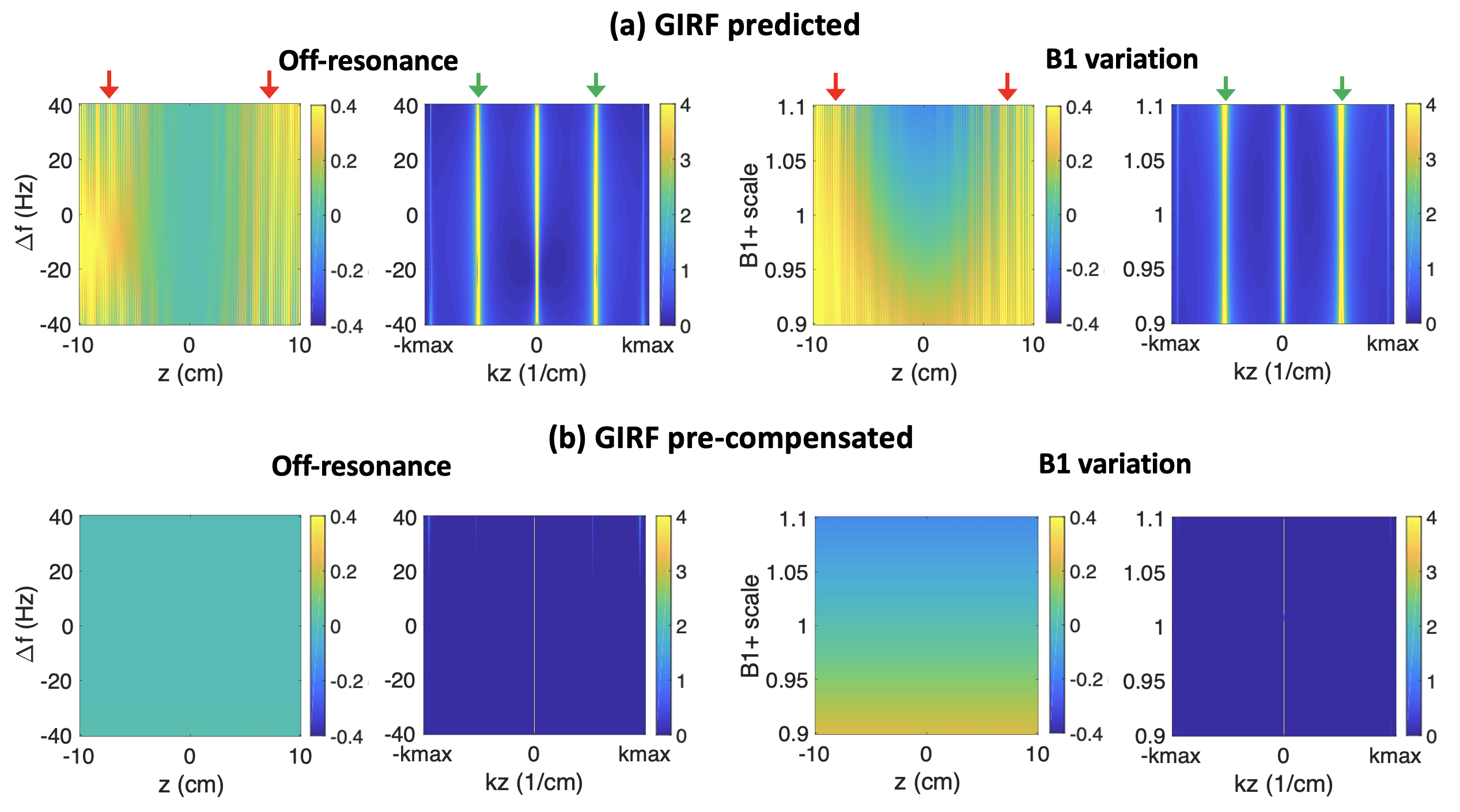
Figure 4. Impact of gradient distortions and pre-compensation. (a) GIRF predicted excitation profiles as a function of ∆f and B1+ scale. (b) GIRF pre-compensated profiles. Notice that stripe artifacts appear as spatial modulations (red arrows) and as sidelobe peaks (green arrows) in (a). These are completely resolved in (b) by using GIRF-based pre-compensation.