Dimitrios G. Gkotsoulias1, Riccardo Metere2, Yanzhu Su1, Cornelius Eichner1, Torsten Schlumm1, Roland Müller1, Alfred Anwander1, Toralf Mildner1, Carsten Jäger1, André Pampel1, Catherine Crockford3,4, Roman Wittig3,4, Liran Samuni 4,5, Kamilla Pleh 6, Chunlei Liu7, and Harald E. Möller1
1Max Planck Institute for Human Cognitive and Brain Sciences, Leipzig, Germany, 2Donders Institute for Brain, Cognition and Behaviour, Nijmegen, Netherlands, 3Max Planck Institute for Evolutionary Anthropology, Leipzig, Germany, 4Tai Chimpanzee Project, Centre Suisse de Recherches Scientifiques en Cote d'Ivoire, Abidjan, Cote D'ivoire, 5Department of Human Evolutionary Biology, Harvard University, Cambridge, MA, United States, 6Project Group Epidemiology of Highly Pathogenic Microorganisms, Robert Koch Institute, Berlin, Germany, 7EECS, UC Berkeley, Berkeley, CA, United States
1Max Planck Institute for Human Cognitive and Brain Sciences, Leipzig, Germany, 2Donders Institute for Brain, Cognition and Behaviour, Nijmegen, Netherlands, 3Max Planck Institute for Evolutionary Anthropology, Leipzig, Germany, 4Tai Chimpanzee Project, Centre Suisse de Recherches Scientifiques en Cote d'Ivoire, Abidjan, Cote D'ivoire, 5Department of Human Evolutionary Biology, Harvard University, Cambridge, MA, United States, 6Project Group Epidemiology of Highly Pathogenic Microorganisms, Robert Koch Institute, Berlin, Germany, 7EECS, UC Berkeley, Berkeley, CA, United States
Orientation-dependent information may be integrated into quantitative susceptibility mapping by employing diffusion-weighted MRI and machine learning.
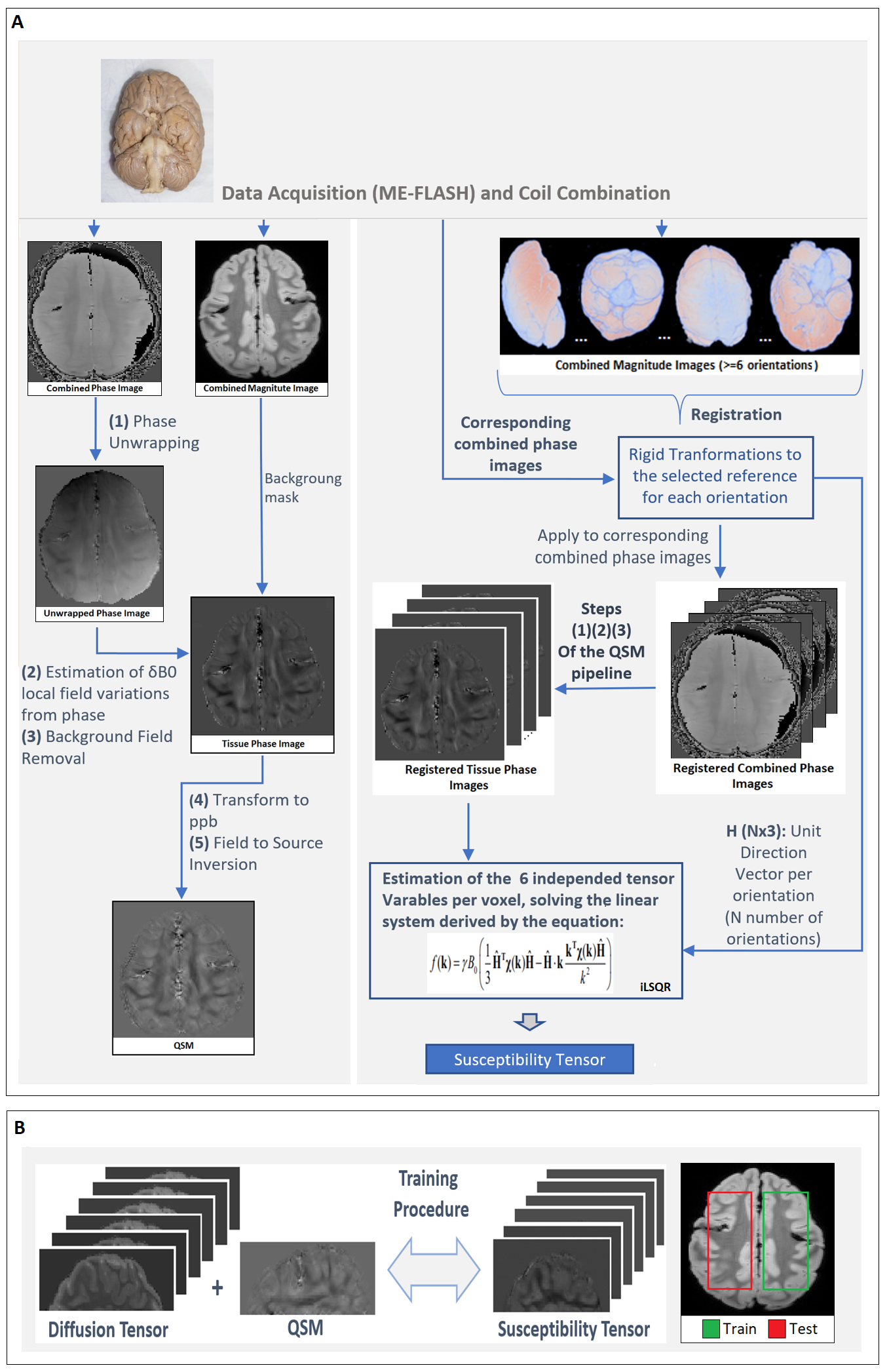
Figure 1. (A) Schematic of the QSM and STI pipelines. The brain used in for the data acquisitions of this abstract comes from a wild, female Chimpanzee (Pan troglodytes verus) named “Emma”, 6 years old, from Tai National Forest, Ivory Coast, West Africa. Cause of death was an attack by another group of Chimpanzees. (B) The general pipeline used for training of the machine learning model.
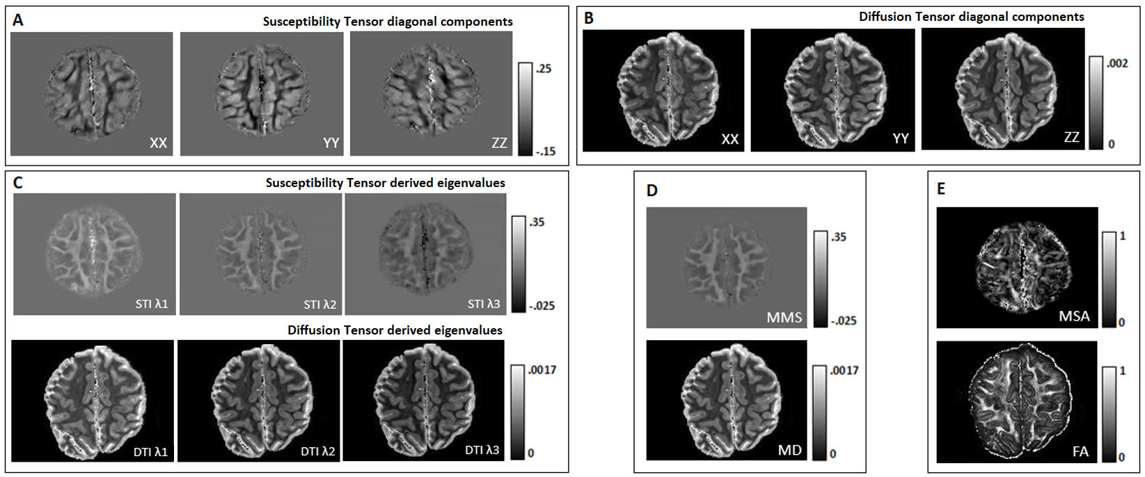
Figure 2. The three
main tensor components (diagonal) from STI (A) and DTI (B). Differentiation of GM/WM
is clear in both tensors. (C) Eigenvalues derived by susceptibility (sign is inverted) and diffusion tensors. (D) Maps
of mean diffusivity (MD) and mean magnetic susceptibility (MMS). (E) Maps of fractional anisotropy (FA) and magnetic susceptibility anisotropy (MSA). MSA exhibits some common spatial characteristics with FA in white matter but with noticeably more variations.