Hyeong-Geol Shin1, Young Hyun Yun2, Seong Ho Yoo2, Sehoon Jung3, Sunhye Kim3, Hyung Jin Choi 2, and Jongho Lee1
1Seoul National University, Seoul, Korea, Republic of, 2Seoul National University College of Medicine, Seoul, Korea, Republic of, 3Research Institute of Industrial Science and Technology, Pohang, Korea, Republic of
1Seoul National University, Seoul, Korea, Republic of, 2Seoul National University College of Medicine, Seoul, Korea, Republic of, 3Research Institute of Industrial Science and Technology, Pohang, Korea, Republic of
The magnetic susceptibility source separation method successfully reconstructs the individual distribution of paramagnetic and diamagnetic sources in Monte-Carlo simulation and delineates anatomical features of iron and myelin histology in the ex-vivo and in-vivo brain.
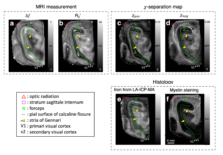
Figure 3. Comparison between ex-vivo 𝜒-separation results vs. iron and myelin histology. (a-b) Frequency shift and R2' maps. (c-d) Positive and negative susceptibility maps from 𝜒-separation. (e) Iron distribution from LA-ICP-MS. (f) Myelin images from LFB staining. Similar features are observed between positive susceptibility map vs. iron image as well as negative susceptibility map vs. myelin image. 𝜒-separation maps also delineate brain anatomy observed in iron and myelin histology (see color markers).
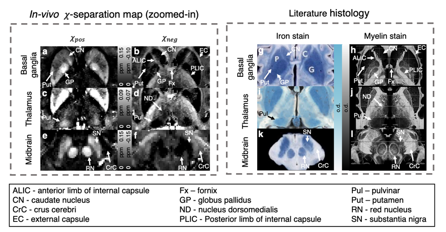
Figure 5. Comparison between in-vivo 𝜒-separation results (a-f) vs. iron and myelin histology from literatures (g-l). The positive and negative susceptibility maps demonstrate similar distributions with histologically stained iron and myelin images, respectively. The 𝜒-separation maps render exquisite details of the brain anatomy that agree with the iron and myelin histology (white arrows).