Kwok-Shing Chan1, Jeroen Mollink2, Jenni Schulz1, Anne-Marie van Cappellen van Walsum2, and José P. Marques1
1Donders Institute for Brain, Cognition and Behaviour, Radboud University, Nijmegen, Netherlands, 2Department of Medical Imaging, Anatomy, Donders Institute for Brain, Cognition and Behaviour, Radboud University Medical Center, Nijmegen, Netherlands
1Donders Institute for Brain, Cognition and Behaviour, Radboud University, Nijmegen, Netherlands, 2Department of Medical Imaging, Anatomy, Donders Institute for Brain, Cognition and Behaviour, Radboud University Medical Center, Nijmegen, Netherlands
Though similar isotropic magnetic susceptibility to in vivo imaging can be derived using formalin-fixed tissue, non-susceptibility contributions such as microstructure effect on MRI phase image is significantly different on fixed tissue.
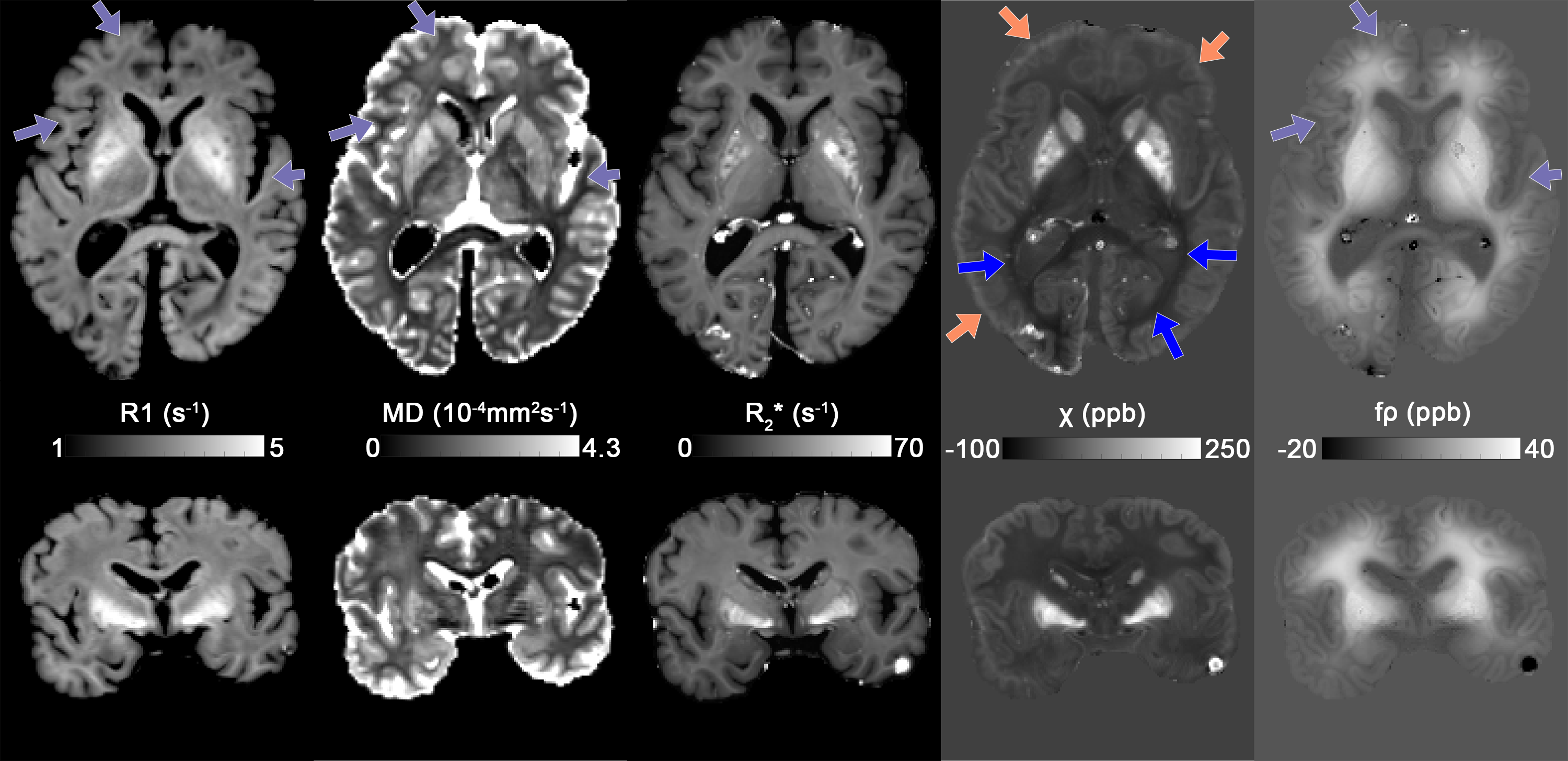
Fig. 2: Transverse and coronal slices of quantitative R1, MD, R2*, χ and fρ maps after fixation. Unlike in vivo, R1 maps show enhanced values in iron-rich DGM, similar to R2* and χ, and greatly reduced GM & WM contrast and overall increased rates. MD in WM is much lower than typical in-vivo values (5-7x10⁻⁴mm2/s). QUASAR χ shows fixation artefact at the brain surface (bright ring, orange arrows). The non-susceptibility source map contrast is dominated by fixation artefact (gradient change from brain surface toward the centre), which are also visible in R1 and MD maps (purple arrows).
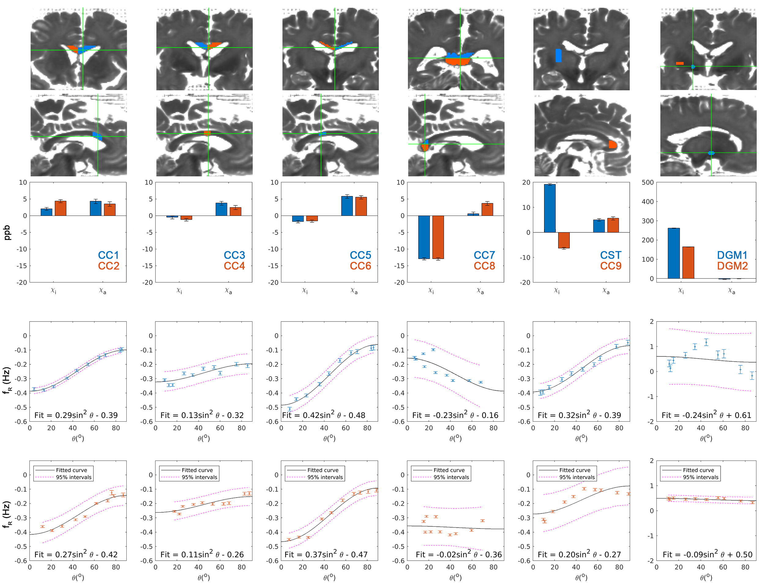
Fig. 3: Top 2 rows show ROIs of excised samples. 3rd row shows χi and χa of excised specimens measured by external field. Bottom 2 rows show residual field inside the sample fitted with Eq.4. WM at the genu and splenium of the corpus callosum (CC7-9) showed both the strongest diamagnetic isotropic susceptibility but very poorly described residual fields. The CST sample had residual DGM tissue in the corner of the sample, explaining the strong positive susceptibility compared to other WM. These 4 samples were excluded from the correlation analysis in Fig.4.