Lin Chen1,2, Anja Soldan3, Kenichi Oishi1, Andreia Faria1, Marilyn Albert3, Peter van Zijl1,2, and Xu Li1,2
1Department of Radiology and Radiological Sciences, Johns Hopkins University, Baltimore, MD, United States, 2F.M. Kirby Research Center for Functional Brain Imaging, Kennedy Krieger Institute, Baltimore, MD, United States, 3Department of Neurology, Johns Hopkins University, Baltimore, MD, United States
1Department of Radiology and Radiological Sciences, Johns Hopkins University, Baltimore, MD, United States, 2F.M. Kirby Research Center for Functional Brain Imaging, Kennedy Krieger Institute, Baltimore, MD, United States, 3Department of Neurology, Johns Hopkins University, Baltimore, MD, United States
Greater volume of multiple brain regions was strongly associated with the rate of cognitive decline. Associations between brain iron and β-amyloid and longitudinal cognitive decline were weaker, with brain iron in the basal ganglia and entorhinal cortex predicting global decline.
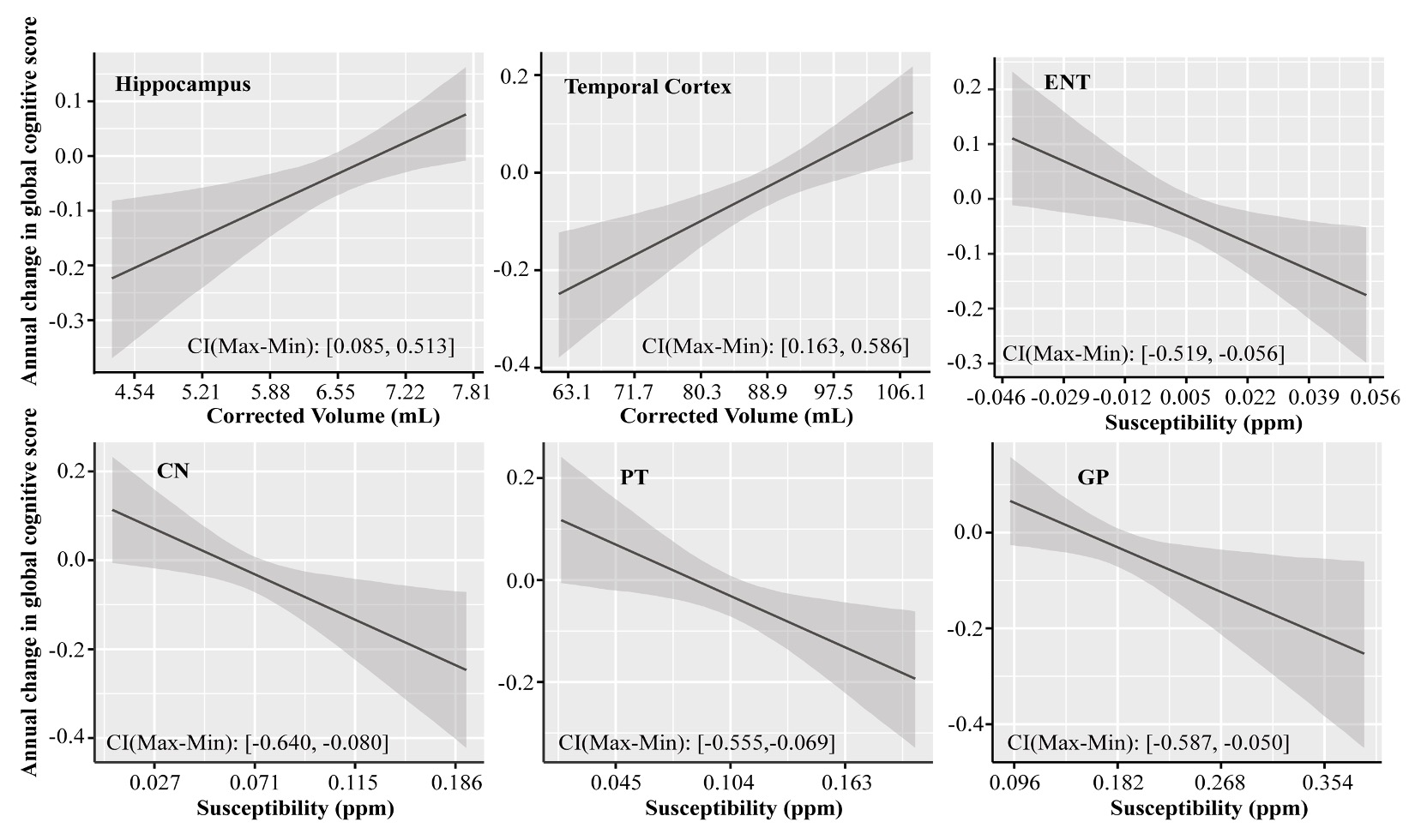
Figure 1 Associations of the corrected volume in hippocampus and temporal cortex, QSM in ENT, CN, PT and GP, to the annual change in global cognitive functions in all participants (n = 150). Annual change of cognitive function was calculated for each participant based on his or her trajectory of decline in the follow-up years after baseline MRI (up to 5 years), and it was plotted against baseline predictor, i.e. corrected volume or QSM. CI (Max-Min) = confidence interval of the difference between the annual change in global cognitive score at the minimum and maximum values of the predictor.
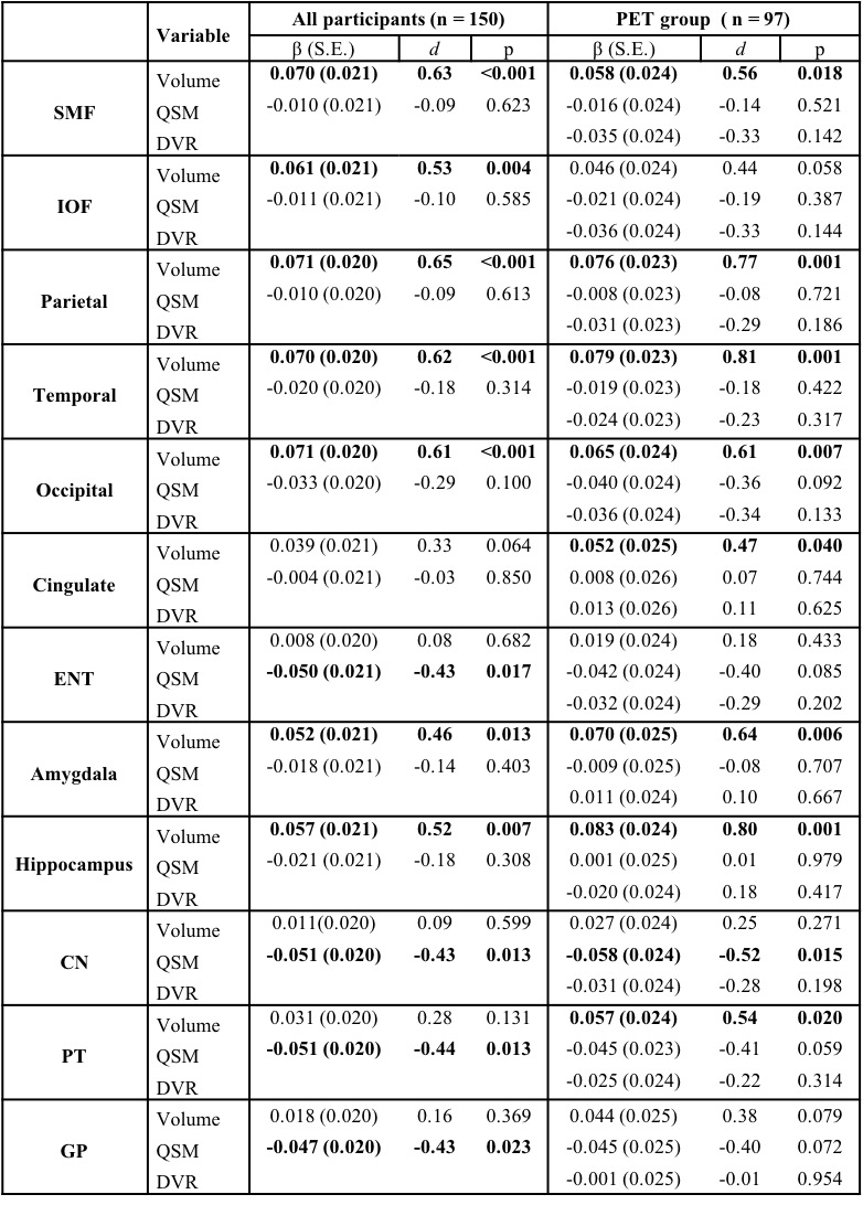
Table 1 Associations of structure volume, tissue iron load (QSM) and β-amyloid load (PET DVR) with rate of change in global cognitive composite scores (variable×time interaction in mixed-effects models) in all participants and participants in the PET group. d: effect size.