Ibrahim Khormi1,2, Oun Al-iedani1,2, Amir Fazlollahi2,3, Bryan Paton2,4, Jeannette Lechner-Scott2,4,5, Abdulaziz Alshehri1,2, Kieran O'Brien6,7, Steffen Bollmann8, Rishma Vidyasagar9, Scott Ayton9, Anne-Louise Ponsonby9,10, and Saadallah Ramadan1,2
1School of Health Sciences, University of Newcastle, Newcastle, Australia, 2Hunter Medical Research Institute, Newcastle, Australia, 3CSIRO Health and Biosecurity, Brisbane, Australia, 4University of Newcastle, Newcastle, Australia, 5John Hunter Hospital, Newcastle, Australia, 6Siemens Healthcare Pty Ltd, Brisbane, Austria, 7ARC Training Centre for Innovation in Biomedical Imaging Technology, The University of Queensland, Brisbane, Australia, 8The University of Queensland, Brisbane, Australia, 9The Florey Institute of Neuroscience & Mental Health, Parkville, Australia, 10Murdoch Children's Research Institute, Royal Children's Hospital, University of Melbourne, Melbourne, Australia
1School of Health Sciences, University of Newcastle, Newcastle, Australia, 2Hunter Medical Research Institute, Newcastle, Australia, 3CSIRO Health and Biosecurity, Brisbane, Australia, 4University of Newcastle, Newcastle, Australia, 5John Hunter Hospital, Newcastle, Australia, 6Siemens Healthcare Pty Ltd, Brisbane, Austria, 7ARC Training Centre for Innovation in Biomedical Imaging Technology, The University of Queensland, Brisbane, Australia, 8The University of Queensland, Brisbane, Australia, 9The Florey Institute of Neuroscience & Mental Health, Parkville, Australia, 10Murdoch Children's Research Institute, Royal Children's Hospital, University of Melbourne, Melbourne, Australia
QSM metrics in caudate showed strong correlations with depression scores,
while pallidum and thalamus correlated significantly with anxiety.
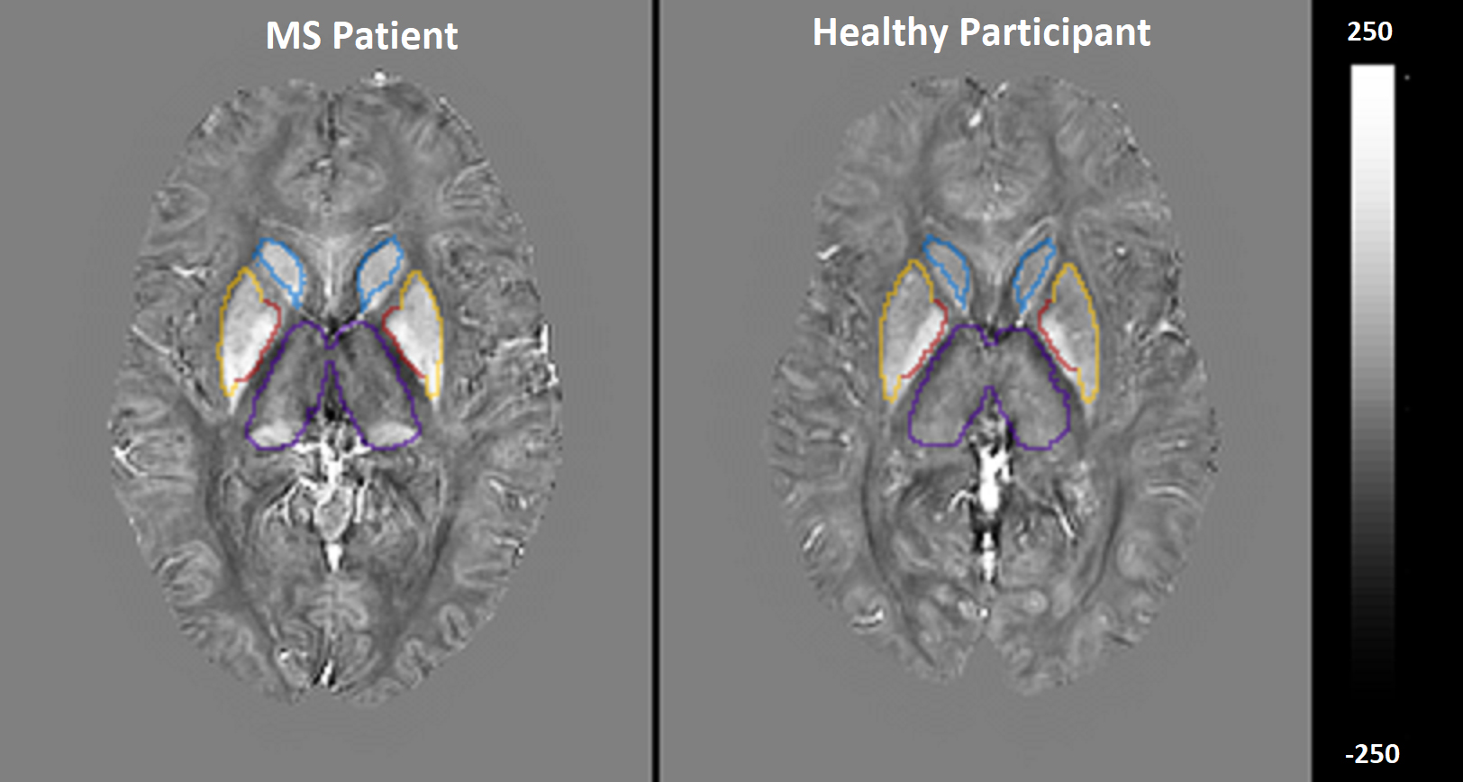
Figure 1. Quantitative
susceptibility maps within thalamus (purple), caudate (blue), pallidum (red)
and putamen (yellow) of a 38 years-old female RRMS vs. 40years-old female HCs.
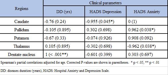
Table 3. Spearman’s correlation between iron-rich regions in deep grey matter
in MS cohort only.