Julian Emmerich1,2, Frederik L. Sandig3, and Sina Straub1
1Division of Medical Physics in Radiology, German Cancer Research Center (DKFZ), Heidelberg, Germany, 2Faculty of Physics and Astronomy, University of Heidelberg, Heidelberg, Germany, 3Division Radiology, German Cancer Research Center (DKFZ), Heidelberg, Germany
1Division of Medical Physics in Radiology, German Cancer Research Center (DKFZ), Heidelberg, Germany, 2Faculty of Physics and Astronomy, University of Heidelberg, Heidelberg, Germany, 3Division Radiology, German Cancer Research Center (DKFZ), Heidelberg, Germany
It is
shown that the occurrence of bright multiple sclerosis lesions and have
different origins that can either be separately observed in positive or
negative susceptibility maps.
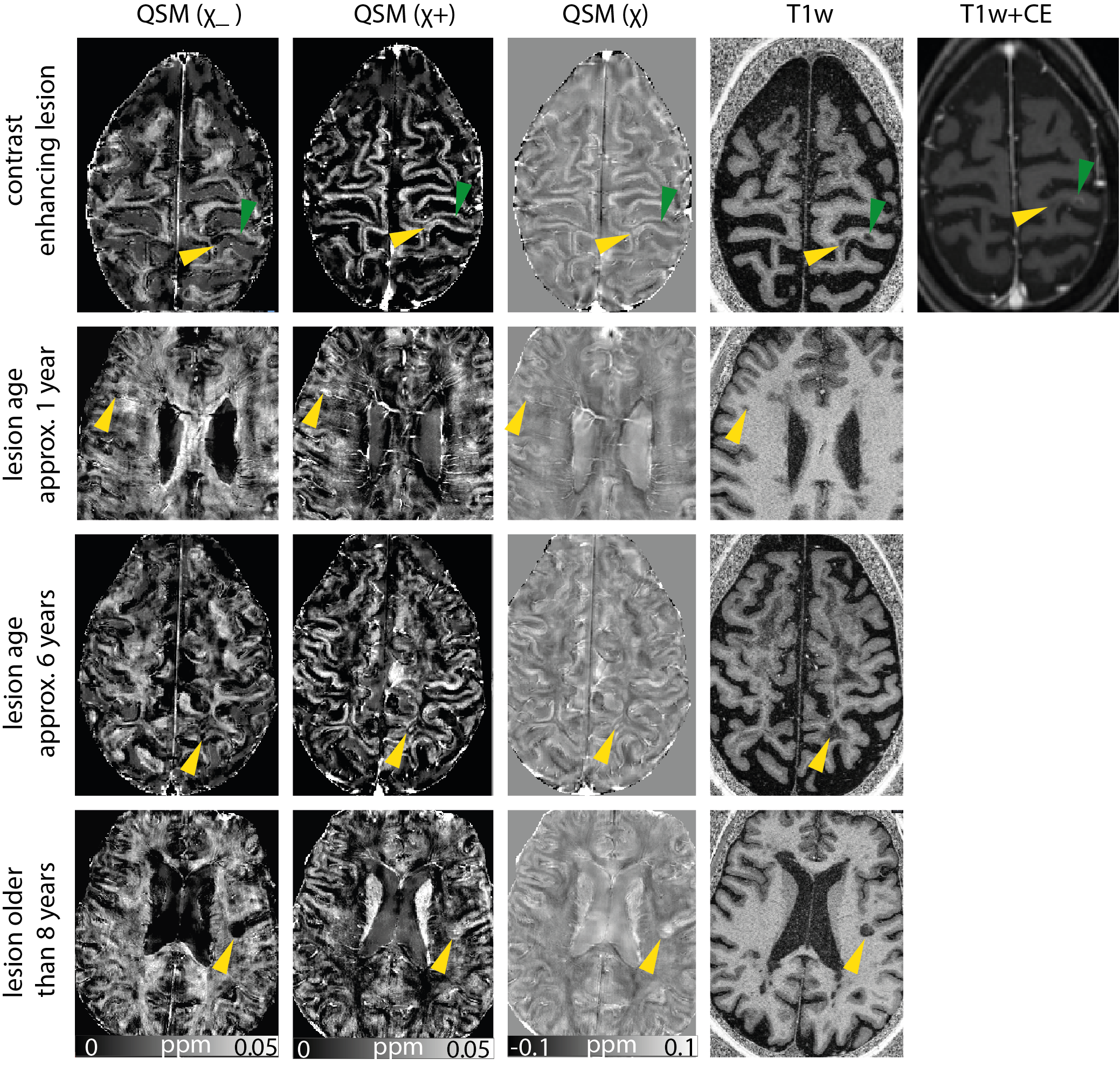
Figure 2:
Positive,
negative and conventional susceptibility maps, as well as a T1-weighted image
for lesions from three different patients. For the enhancing lesion, additionally
a contrast-enhanced T1-weighted image is shown. Lesion age is indicated on the
left and lesions are ordered according to their age. Green indicates a contrast
enhancing lesion, yellow arrows point to non-enhancing lesions.
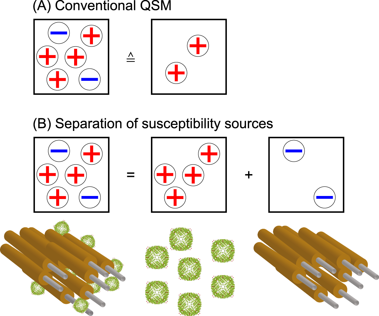
Figure1: The cancellation of the susceptibility effects within the same voxel in
QSM are illustrated.