Alexander Mark Weber1, Yuting Zhang2, Christian Kames3, and Alexander Rauscher1
1Pediatrics, UBC, Vancouver, BC, Canada, 2Radiology, Children’s Hospital of Chongqing Medical University, Chongqing, China, 3Physics, UBC, Vancouver, BC, Canada
1Pediatrics, UBC, Vancouver, BC, Canada, 2Radiology, Children’s Hospital of Chongqing Medical University, Chongqing, China, 3Physics, UBC, Vancouver, BC, Canada
QSM derived oxygen saturation values in healthy term and asphixia injured preterm and term neonates agrees well with past literature. CSVO2 in preterm and term neonates with HIE, however, were not found to be significantly different from each other or healthy controls.
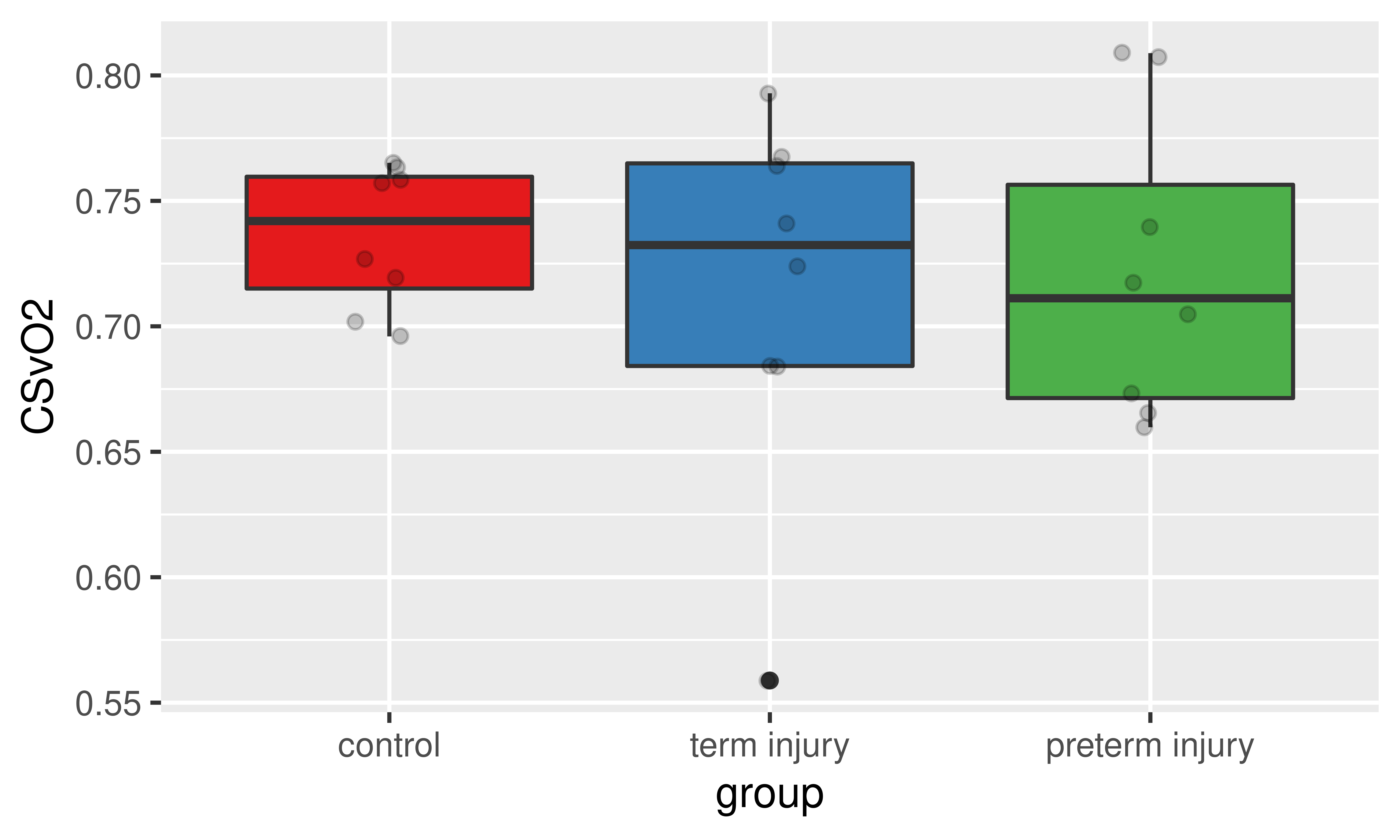
Figure 2. Boxplot of CSVO2 percentages by group. Grey circles are the ROI measurements from each subject.
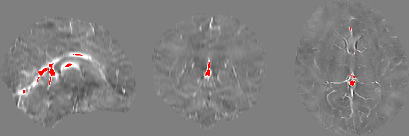
Figure 1. Sample internal veins selected after thresholding out the lower 99.75% χ values. As can be seen in these sagittal, coronal, and axial views from a sample healthy term neonate, the major veins that were left over include the straight sinus, inferior sagittal sinus and the internal cerebral vein. Note the weak contrast between grey and white matter and the basal ganglia due to the low myelin and iron content of the newborn brain.