Russell Murdoch1, Hanne Stotesbury2, Jamie Kawadler2, Dawn Saunders2, Fenella Kirkham2, and Karin Shmueli1
1Department of Medical Physics and Biomedical Engineering, University College London, London, United Kingdom, 2Imaging and Biophysics, Developmental Neurosciences, UCL Great Ormond Street Institute of Child Health, London, United Kingdom
1Department of Medical Physics and Biomedical Engineering, University College London, London, United Kingdom, 2Imaging and Biophysics, Developmental Neurosciences, UCL Great Ormond Street Institute of Child Health, London, United Kingdom
Silent cerebral infarcts in the parietal lobes of the brain had significantly lower magnetic susceptibility than frontal lobe lesions. Strong correlations were observed between lesion susceptibilities measured using two different GRE acquisitions suggesting χ referencing is not needed.
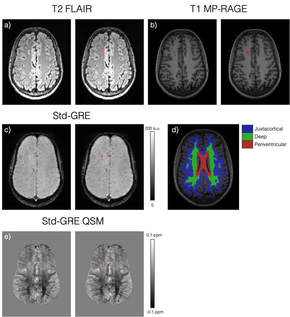
a) Axial slice of a T2-weighted FLAIR image showing two silent cerebral infarcts (SCI) in a representative sickle cell anaemia subject (right: SCI ROI overlaid in red). b) Axial slice of the T1-weighted MP-RAGE image showing the same lesions appearing hypointense. c) Similar axial slice of the Std-GRE magnitude at TE = 27ms in the same subject. d) A different axial slice from the MP-RAGE showing the segmented juxtacortical, deep and periventricular white matter regions. e) QSM in the same axial slice as in c) showing a slight χ difference between the lesions and normal appearing WM.
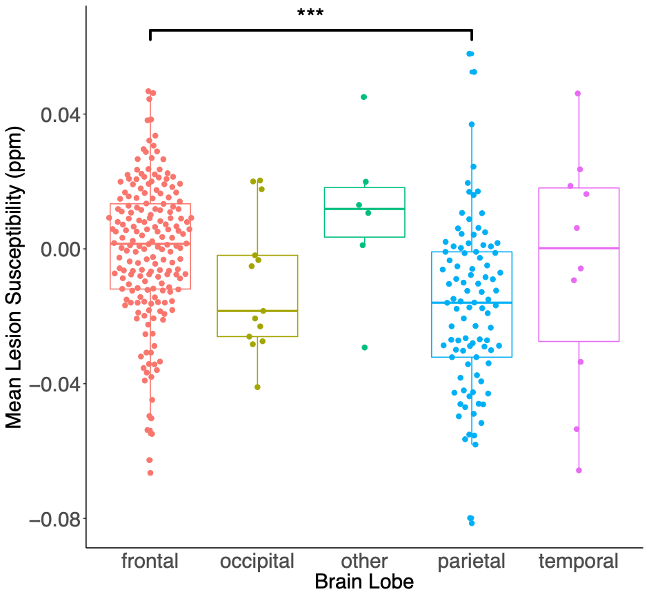
Mean SCI lesion χ within each brain lobe in the combined sickle cell anaemia and healthy control groups. Mean SCI χ are significantly lower for lesions in the parietal lobe relative to the frontal lobe (diff = -0.017ppm, p<0.001). In white matter, myelin is the key diamagnetic (negative) susceptibility source. Therefore, these results suggest reduced myelin concentrations within lesions in the frontal lobe relative to the parietal lobe. No significant χ differences were observed between SCI any other regions.
