Ashmita De1, Derek J. Emery2, Kenneth S. Butcher3, and Alan H. Wilman1
1Department of Biomedical Engineering, University of Alberta, Edmonton, AB, Canada, 2Department of Radiology and Diagnostic Imaging, University of Alberta, Edmonton, AB, Canada, 3Division of Neurology, Department of Medicine, University of Alberta, Edmonton, AB, Canada
1Department of Biomedical Engineering, University of Alberta, Edmonton, AB, Canada, 2Department of Radiology and Diagnostic Imaging, University of Alberta, Edmonton, AB, Canada, 3Division of Neurology, Department of Medicine, University of Alberta, Edmonton, AB, Canada
tSWI removes the phase artifacts that are observed in SWI, provides
better susceptibility weighting than magnitude and improves contrast visualization
within the hemorrhage as compared to QSM. tSWI can be used for hemorrhage
visualization when SWI cannot provide a clear depiction.
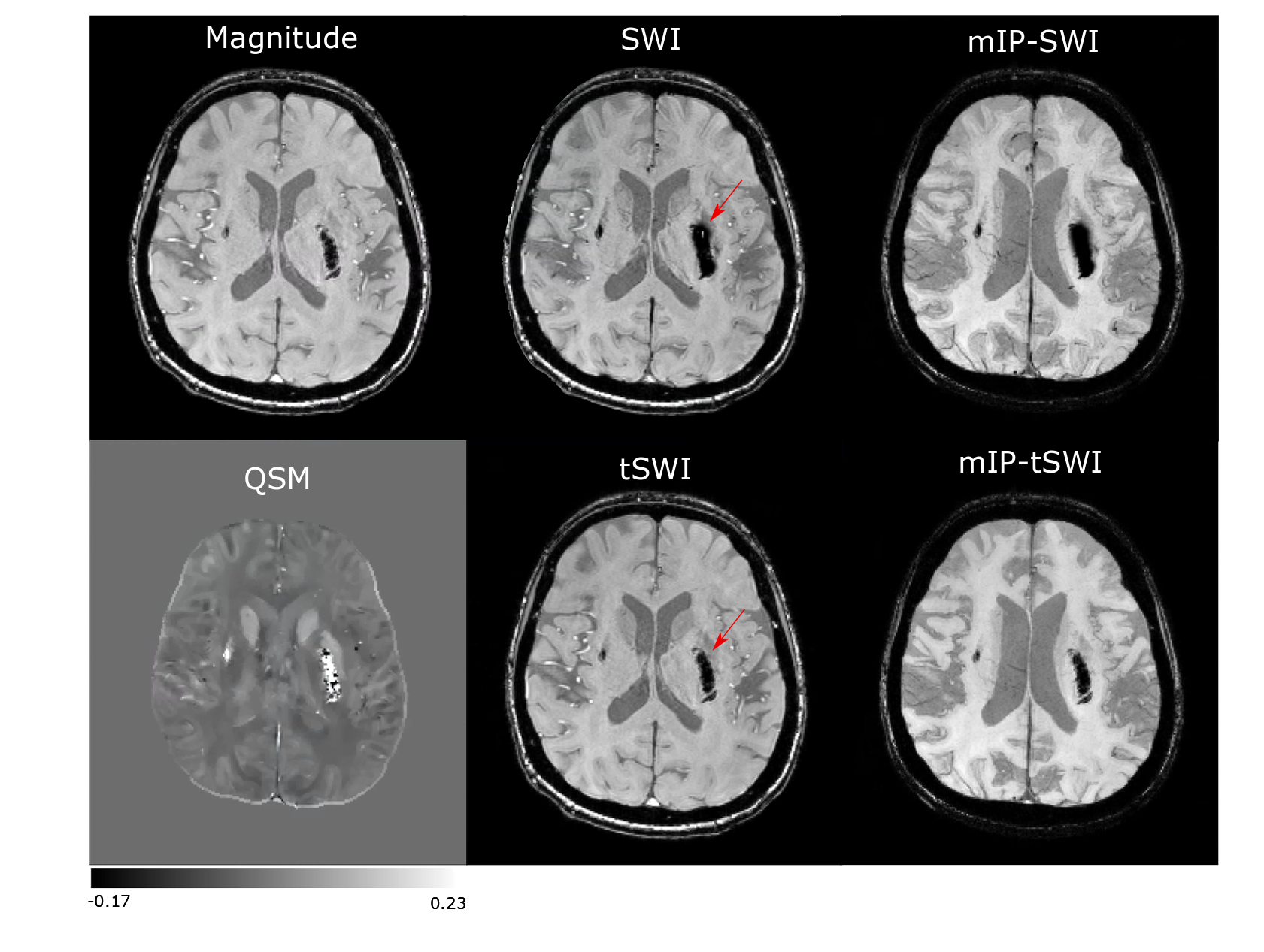
Figure 1: Magnitude, SWI,
tSWI, QSM and mIP from SWI and tSWI in a patient with an acute Day 2 hemorrhage. It demonstrates the blooming effect in SWI of hemorrhage.
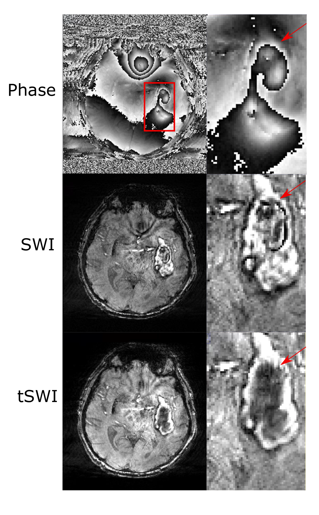
Figure 2: A comparison between phase, SWI and tSWI in a patient with Day 12 hemorrhage to show phase wrap artifacts are present in SWI for large hemorrhage which gets removed in tSWI as shown by the red arrow.
