Marjolein Bulk1, Thijs van Harten1, Boyd Kenkhuis1, Francesca Inglese1, Ingrid Hegeman1, Sjoerd van Duinen1, Ece Ercan1, Cesar Magro-Checa1,2, Jelle Goeman1, Christian Mawrin3, Mark van Buchem1, Gerda Steup-Beekman1, Tom Huizinga1, Louise van der Weerd1, and Itamar Ronen1
1Leiden University Medical Center, Leiden, Netherlands, 2Zuyderland Medical Center, Heerlen, Netherlands, 3Otto-von-Guericke University, Magdeburg, Germany
1Leiden University Medical Center, Leiden, Netherlands, 2Zuyderland Medical Center, Heerlen, Netherlands, 3Otto-von-Guericke University, Magdeburg, Germany
No significant
differences in local susceptibility in the basal ganglia were found between SLE
patients with NP complaints and healthy controls, suggesting that in NPSLE, neuroinflammation
is not necessarily associated with iron accumulation.
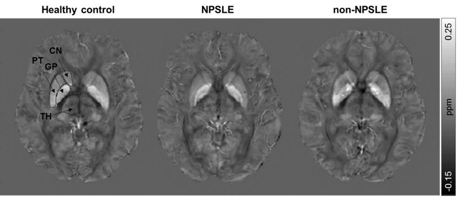
Figure 2. Representative axial QSM images of a
healthy control, NPSLE patient and non-NPSLE patient. The regions of interest are
indicated in the healthy control. TH = Thalamus; CN = Caudate nucleus; PT =
Putamen; GP = Globus pallidus.
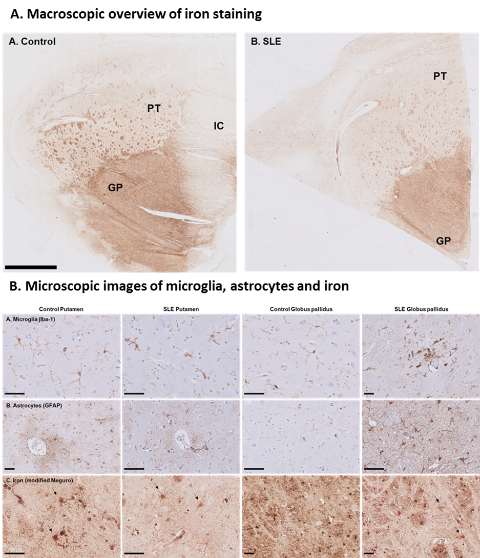
Figure 5. Iron in control and SLE brain. Globus pallidus showed higher
staining intensity compared to putamen. Within the putamen small areas of increased
iron were found originating from myelinated fiber bundles. Iron was
predominantly found in oligodendrocytes and myelin, to a lesser extent, in
neurons, microglia and astrocytes (arrows) in both control and SLE.