Lee-Ren Yeh1, Yang Zhang2, Jeon-Hor Chen2, An-Chi Wang3, JieYu Yang3, Peter Chang2, Daniel Chow2, and Min-Ying Su2
1Radiology, E-Da Hospital, Kaohsiung, Taiwan, 2University of California Irvine, Irvine, CA, United States, 3Radiology, Chi-Mei Medical Center, Tainan, Taiwan
1Radiology, E-Da Hospital, Kaohsiung, Taiwan, 2University of California Irvine, Irvine, CA, United States, 3Radiology, Chi-Mei Medical Center, Tainan, Taiwan
Deep learning using ResNet50 for differentiating
malignant from benign vertebral fracture achieved a satisfactory diagnostic
accuracy of 92%, although inferior to 98% made by a senior MSK radiologist, was
much higher compared to 66% made by a R1 resident.
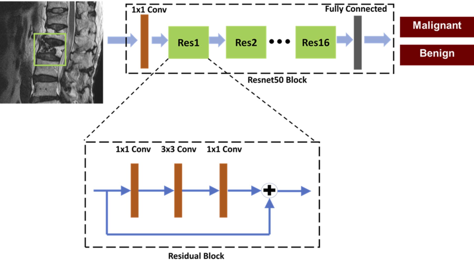
Figure 1. Architecture of
ResNet50, containing 16 residual blocks. Each residual block begins with one
1x1 convolutional layer, followed by one 3x3 convolutional layer and ends with
another 1x1 convolutional layer. The output is then added to the input via a
residual connection. The total input number is 6: T1W and T2W of the slice with
its two neighboring slices, so one convolutional layer with 1x1 filter is added
before ResNet to extract interchannel features and transform from 6 channels to
3 channels as input.
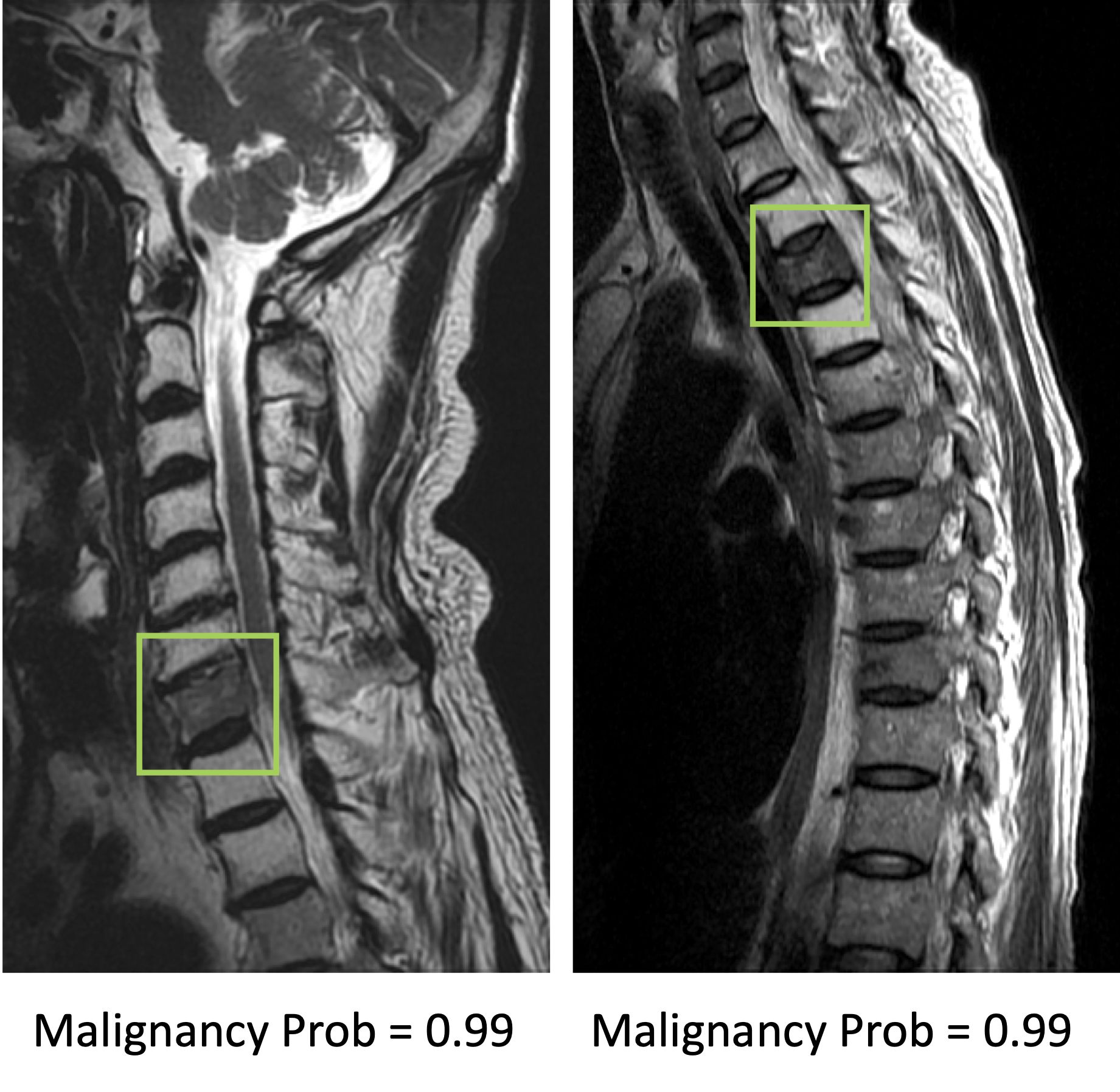
Figure 2. Two true positive
malignant cases. The image at left panel shows diffuse tumor infiltration at
the 7th cervical (C7) vertebral body with posterior cortical destruction and no
apparent collapse. The image at right panel shows diffuse tumor infiltration at
third thoracic (T3) vertebra with anterior wedge deformity. The
fatty change of other cervical vertebrae in the left panel and T2/T4 vertebrae
in right panel is post-radiation effect.