Emma Bahroos1, Misung Han1, Cynthia Chin1, David Shin2, Javier Villanueva-Meyer1, Thomas Link1, Valentina Pedoia1, and Sharmila Majumdar1
1Radiology and Biomedical Imaging, University of California San Francisco, San Francisco, CA, United States, 2Applications and Workflow, GE Healthcare, Menlo Park, CA, United States
1Radiology and Biomedical Imaging, University of California San Francisco, San Francisco, CA, United States, 2Applications and Workflow, GE Healthcare, Menlo Park, CA, United States
Our results show that scan times can be cut in half using a reduced NEX protocol and loss of SNR can be recovered by a DL image reconstruction algorithm, without a severe degradation in the ability to discern anatomical structures. A potential tool for faster imaging in patients with severe LBP.
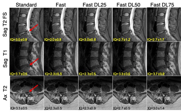
Figure 2: Comparing images from the standard and fast acquisitions and DL-reconstructed images (Standard, Fast, Fast DL25, Fast DL50, Fast DL75 images). The mean scores from the three radiologists for the ‘overall image quality’ is stated on each image. Large disc protrusion is depicted with the arrow on each sequence.
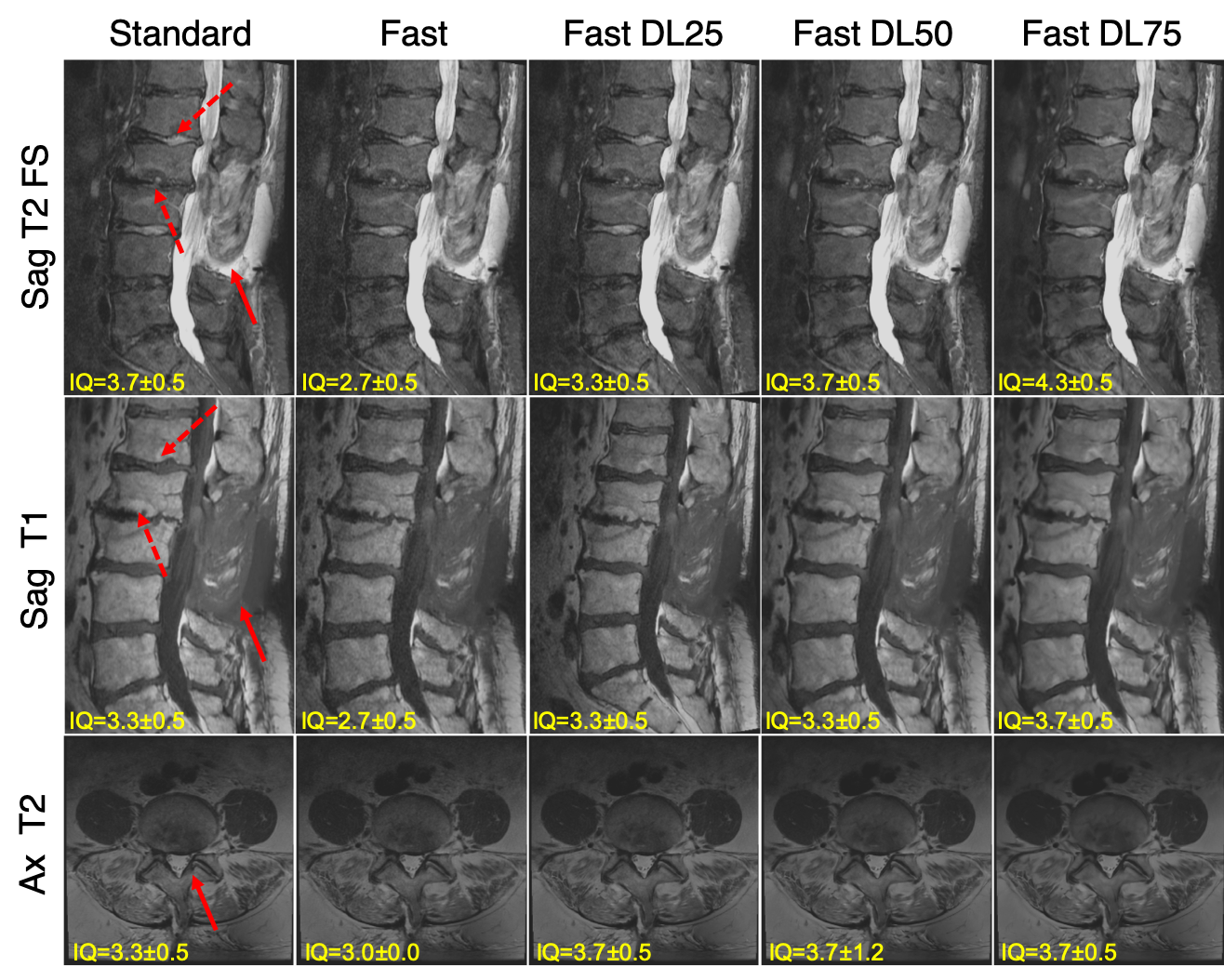
Figure 1: Comparing images from the standard and fast acquisitions and DL-reconstructed images (Standard, Fast, Fast DL25, Fast DL50, Fast DL75 images). The mean scores from the three radiologists for the ‘overall image quality’ is stated on each image. Changes in multiple vertebral bone marrow can be seen on sagittal images (depicted by solid and dashed arrows, respectively), and facet hypertrophy (depicted by an arrow) on axial images.