Weixin He1, Qi Zeng1, Ziwei Zhang1, Xia Zhu1, Zhaoshu Huang2, Lisha Nie3, and Lingling Song1
1Affiliated Hospital of Guizhou Medical University, Guiyang, China, 2592159673@qq.com, Guiyang, China, 3GE Healthcare, MR Research China, Beijing, China
1Affiliated Hospital of Guizhou Medical University, Guiyang, China, 2592159673@qq.com, Guiyang, China, 3GE Healthcare, MR Research China, Beijing, China
The ADC value of bone marrow lesions in OA is higher than that in bone contusions, with a statistical difference. ADC value has the potential value to differentiate the two kinds of lesions.
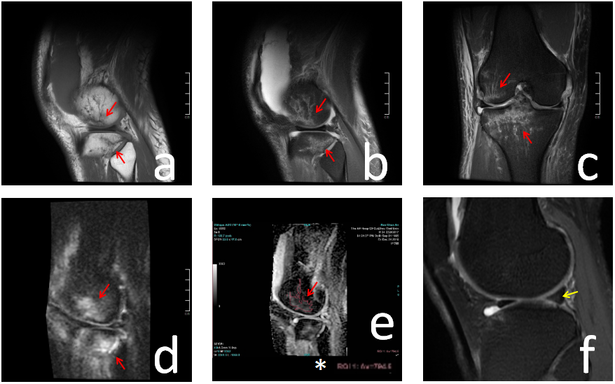
Figure 1 A 24-year-old male suffered multiple injuries caused by traffic accident for more than 3 days. Bone contusions (red arrow) showed striped low signals on T1 sequence (Figure a), while striped high signals on PD-TSE (Figure b-c), DWI (Figure d) and ADC (Figure e). The injury of the medial meniscus (yellow arrow) displayed small patchy high signals on PD-TSE (Figure f). *ADC value of ROI: 794.6 (× 10-6 mm2/S)
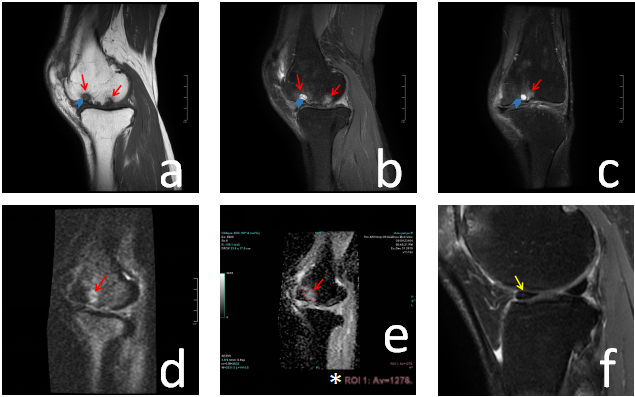
Figure 2 A 62-year-old female suffered from pain of the right knee with no obvious causes, accompanied by limited movement for more than 3 years. The bone marrow lesions of OA (red arrow) showed small patchy and irregular low signals on T1 (Figure a), while small patchy high signals on PD-TSE (Figure b-c), DWI (Figure d) and ADC (Figure e). Cystic degeneration of bone marrow lesions (blue arrow) displayed round-like low signals on T1 (Figure a) while round-like high signals on PD-TSE (Figure b-c). *ADC value of ROI: 1,278 (× 10-6 mm2/S)