Ke Zhang1 and Guobin Hong1
1Radiology, the Fifth Affiliated Hospital, Sun Yat-sen University, Zhuhai, China
1Radiology, the Fifth Affiliated Hospital, Sun Yat-sen University, Zhuhai, China
Synthetic MRI can achieve similar qualitative diagnostic performance to detect sacroiliac joint lesions compared with conventional MRI and could be used for accurate relaxation time quantitative diagnosis, accurately distinguishing BME and fat metaplasia.
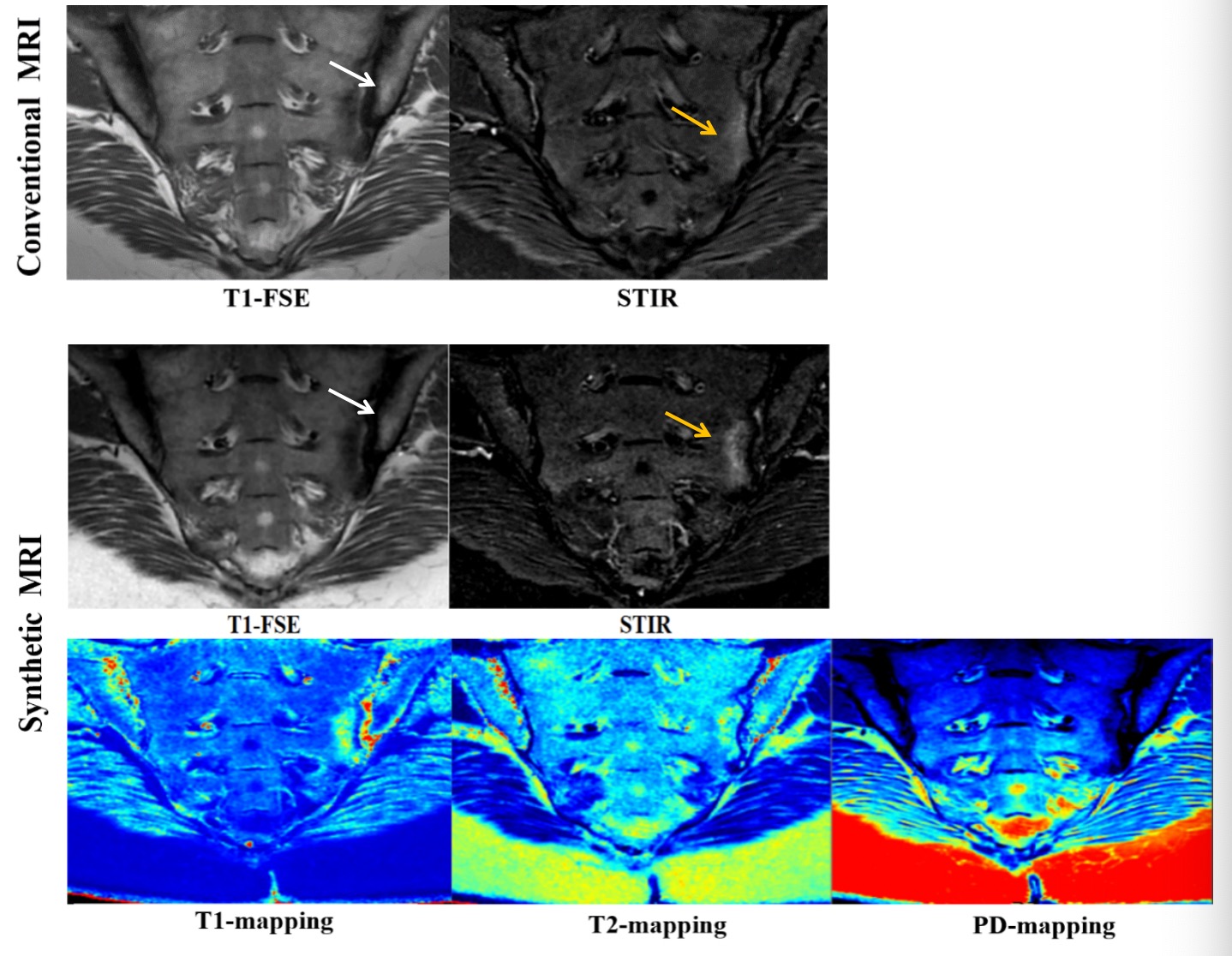
Images in a 38-year-old woman with axSpA.Conventional and synthetic MR images of the sacroiliac joint show the clear presence of BME in the left sacrum articular surface(yellow arrow)that is hyperintense on STIR.The lesion on T1-mapping and T2-mapping show higher value than the surrounding.Conventional and synthetic MR images of the sacroiliac joint show the fat metaplasia in the left iliac articular surface(white arrow)that is hyperintense on T1-FSE.The lesion on T2-mapping and PD-mapping show higher value than the surrounding while T1-mapping shows lower value.