Agazi Samuel Tesfai1, Johannes Fischer1, Ali Caglar Özen1,2, Patrick Eppenberger3, Lena Öhrström3, Frank Rühli3, Ute Ludwig1, and Michael Bock1
1Dept. of Radiology, Medical Physics, Medical Center – University of Freiburg, Faculty of Medicine, University of Freiburg, Freiburg, Germany, 2German Consortium for Translational Cancer Research Partner Site Freiburg, German Cancer Research Center (DKFZ), Heidelberg, Germany, 3Institute of Evolutionary Medicine, Faculty of Medicine, University of Zurich, Zurich, Switzerland
1Dept. of Radiology, Medical Physics, Medical Center – University of Freiburg, Faculty of Medicine, University of Freiburg, Freiburg, Germany, 2German Consortium for Translational Cancer Research Partner Site Freiburg, German Cancer Research Center (DKFZ), Heidelberg, Germany, 3Institute of Evolutionary Medicine, Faculty of Medicine, University of Zurich, Zurich, Switzerland
MR and CT acquisitions of an ancient Child Mummy were compared and combined to increase tissue differentiation.
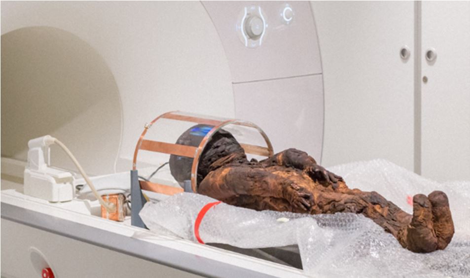
Figure 1: Child
mummy in a custom-built Tx/Rx Birdcage coil with fast switching circuitry connected
to a clinical 3T MRI.
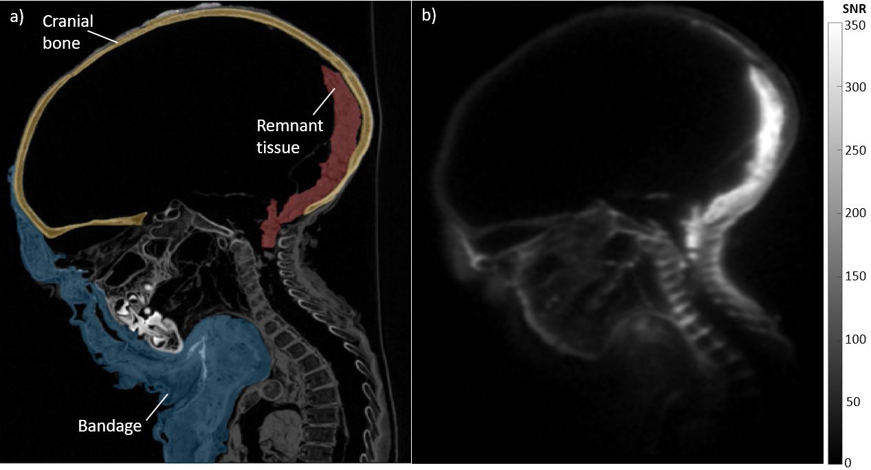
Figure 2: Direct
comparison of the child mummy head images between a) CT image (100 kV) and b) MR
image (TE=70 µs). Signal intensities were evaluated in the marked ROIs.