Miyuki Takasu1, Makoto Iida1, Yasutaka Baba2, Yuji Akiyama1, Yuji Takahashi1, Takashi Abe3, and Kazuo Awai1
1Department of Diagnostic Radiology, Hiroshima University Hospital, Hiroshima, Japan, 2Department of Radiology, International Medical Center, Saitama Medical University, Saitama, Japan, 3Department of Radiology, Nagoya University Hospital, Aichi, Japan, Nagoya, Japan
1Department of Diagnostic Radiology, Hiroshima University Hospital, Hiroshima, Japan, 2Department of Radiology, International Medical Center, Saitama Medical University, Saitama, Japan, 3Department of Radiology, Nagoya University Hospital, Aichi, Japan, Nagoya, Japan
Machine learning with logistic regression model
resulted in the best performance for differentiating MDS from AA using
T1-weighted images. The
model was not predictive for STIR or concatenated images, and performance was
affected by
institutional differences.
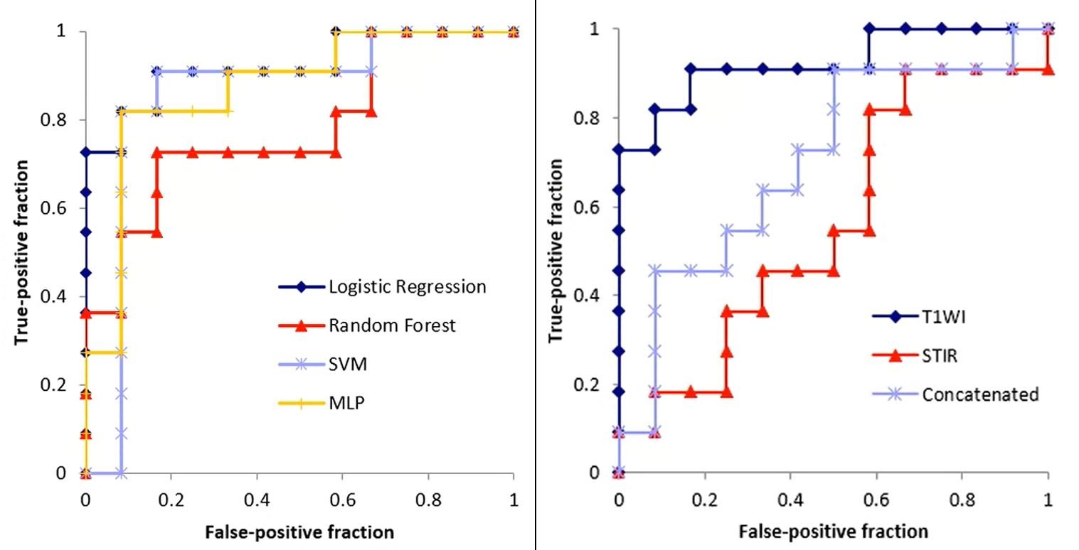
Figure 3. Left: In Scheme 1, the logistic regression (LR) model has the best performance. Receiver-operating characteristic analysis revealed the superior performance of this model for T1-weighted images (AUC, 0.92). Right: The LR model was not predictive for STIR or concatenated images (AUC, 0.56 and 0.71, respectively).
SVM, support vector machine; MLP, multilayer perceptron.
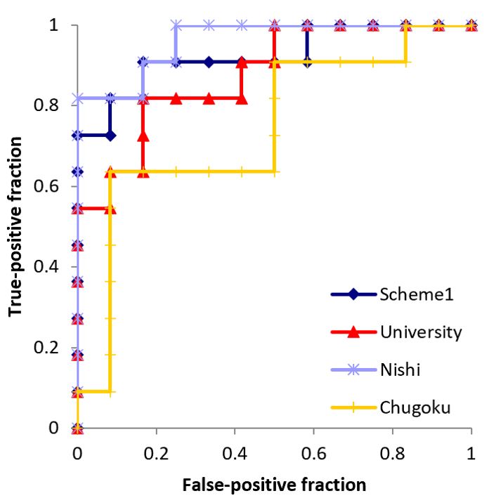
Figure 5. Receiver-operating characteristic curves for differentiating myelodysplastic
syndrome from aplastic anemia using T1-weighted images with Scheme 2. Institutional
differences in performance are apparent (area under the curve: University,
0.879; Nishi, 0.962; Chugoku, 0.742).