Madeleine A. Gao1, Ek T. Tan1, John Neri1, Bin Lin1, Alissa J. Burge1, Hollis G. Potter1, Kevin M. Koch2, and Matthew F. Koff1
1Radiology and Imaging, Hospital of Special Surgery, New York, NY, United States, 2Medical College of Wisconsin, Milwaukee, WI, United States
1Radiology and Imaging, Hospital of Special Surgery, New York, NY, United States, 2Medical College of Wisconsin, Milwaukee, WI, United States
MAVRIC-based DWI shows promise as a biomarker for differentiation of synovial reactions in total hip arthroplasty. Our results display an increased apparent diffusion coefficient for patients with abnormal and fluid/mixed type synovial reactions.
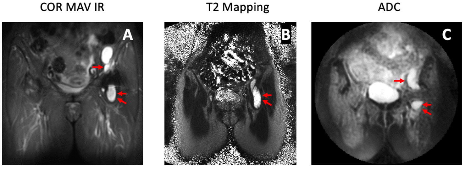
Figure
1. (A) Coronal MAVRIC inversion recovery (IR), (B) T2 mapping,
and (C) apparent diffusion coefficient (ADC) images from a 67 y.o. female
subject with a synovial reaction classified as ‘metallosis’ synovial
impression. Arrows point to regions of interest selected for ADC and T2
mapping analysis.
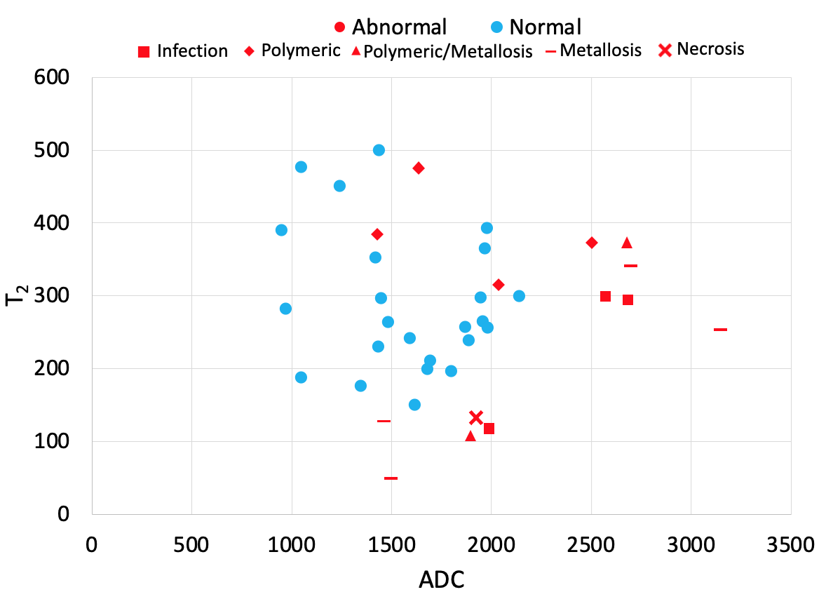
Figure
2. ADC and T2
values for ‘Abnormal’ and ‘Normal’ classifications.