Jay M. Pittman1,2, Aritrick Chatterjee1,2, Teodora Szasz3, Grace Lee1,2, Mihai Giurcanu4, Milica Medved1,2, Ambereen Yousuf1,2, Ajit Devaraj5, Aytekin Oto1,2, and Gregory S. Karczmar1,2
1Radiology, The University of Chicago, Chicago, IL, United States, 2Sanford J. Grossman Center of Excellence in Prostate Imaging and Image Guided Therapy, The University of Chicago, Chicago, IL, United States, 3Research Computing Center, The University of Chicago, Chicago, IL, United States, 4Department of Public Health Sciences, The University of Chicago, Chicago, IL, United States, 5Philips Research North America, Cambridge, MA, United States
1Radiology, The University of Chicago, Chicago, IL, United States, 2Sanford J. Grossman Center of Excellence in Prostate Imaging and Image Guided Therapy, The University of Chicago, Chicago, IL, United States, 3Research Computing Center, The University of Chicago, Chicago, IL, United States, 4Department of Public Health Sciences, The University of Chicago, Chicago, IL, United States, 5Philips Research North America, Cambridge, MA, United States
Standard averaging of multiple acquisitions for high b-values in DWI can obscure cancers, due to very large inter-acquisition variability. We
propose alternatives, including ‘Editing for Restricted Diffusion’, to improve
diagnostic accuracy.
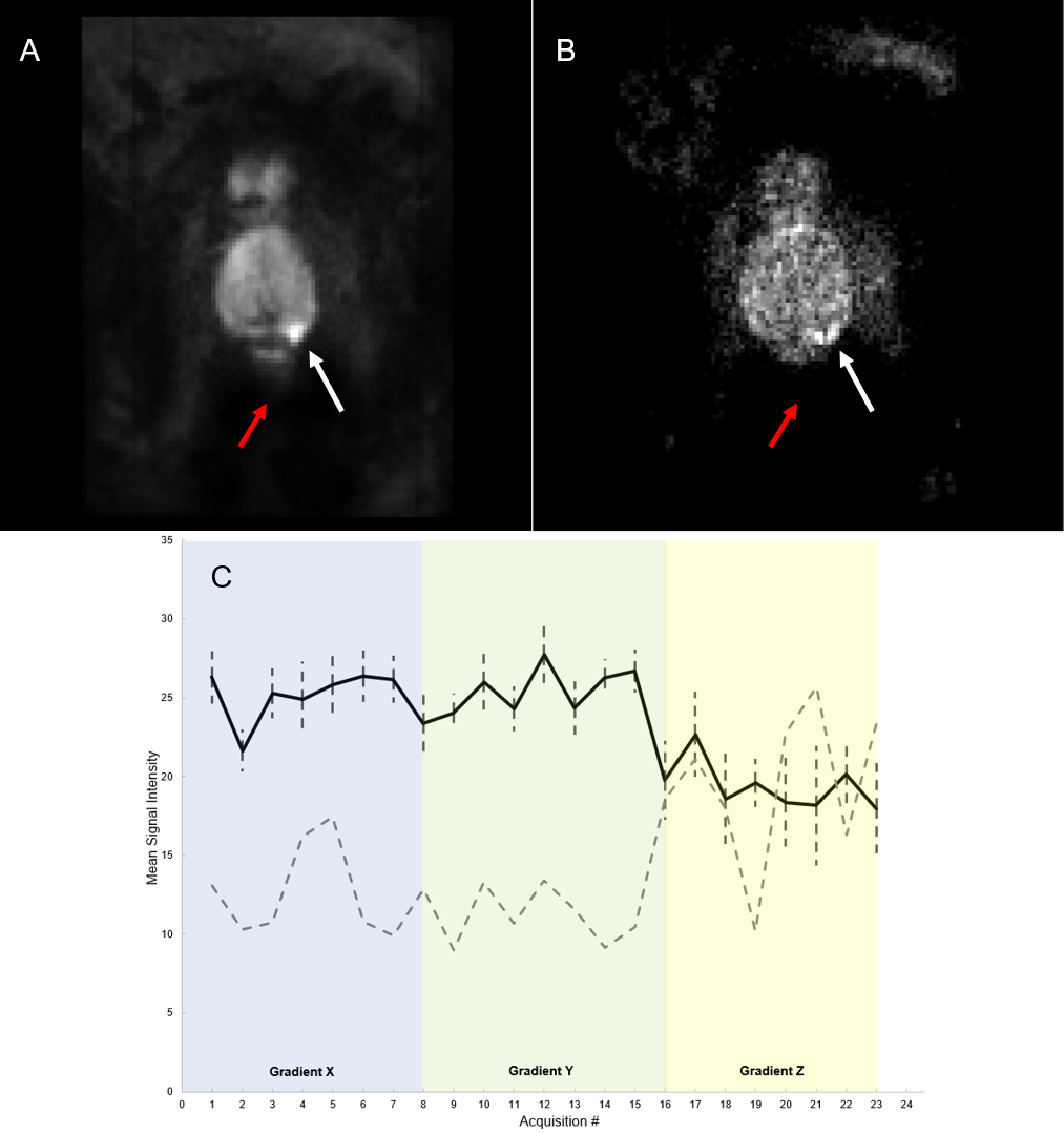
Figure 1. (A) Mean DW Image (900 sec/mm2) of patient 6. (B)
Standard deviation per voxel image for patient 6. (C) Mean signal intensity
versus acquisition # divided into three diffusion-sensitizing gradient
directions (Black line = mean, Dashed error bar = margin of error, Dashed gray
line = range). White arrow points to cancerous lesion while red arrow points to
noise.
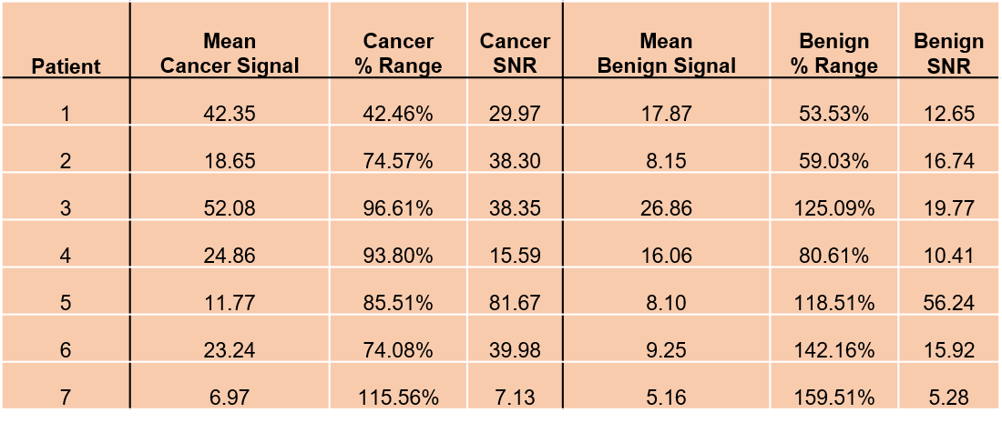
Table 1. Mean Signal, % Range, and SNR of patients 1-7 for a 3x3 region of voxels
in cancerous and benign contralateral tissue.