Kaibao Sun1, Guangyu Dan1,2, Zheng Zhong1,2, and Xiaohong Joe Zhou1,2,3
1Center for MR Research, University of Illinois at Chicago, Chicago, IL, United States, 2Department of Bioengineering, University of Illinois at Chicago, Chicago, IL, United States, 3Departments of Radiology and Neurosurgery, University of Illinois at Chicago, Chicago, IL, United States
1Center for MR Research, University of Illinois at Chicago, Chicago, IL, United States, 2Department of Bioengineering, University of Illinois at Chicago, Chicago, IL, United States, 3Departments of Radiology and Neurosurgery, University of Illinois at Chicago, Chicago, IL, United States
A novel multi-readout
DWI sequence was capable of acquiring multiple diffusion-weighted images at
different TEs in a single scan. This sequence was used to reveal the coupling
between ADC and TE in the healthy and neoplastic prostate tissues.
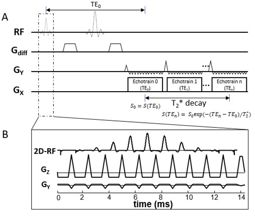
Figure 1: (A): A diagram of the multi-readout
DWI sequence. Multiple EPI echo-trains (or readout trains) are incorporated into
the sequence, with each echo-train corresponding to a specific effective TE. (B):
In the multi-readout DWI sequence, signal excitation is accomplished by a 2D RF
pulse to restrict the FOV so that a shorter echo train length can be used in
each echo-train, allowing multiple echo-trains to be acquired.
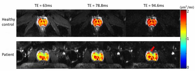
Figure 4: ADC maps of the prostate at
different TEs overlaid onto the corresponding T2*-weighted images (b-value
of 0 s/mm2) of a healthy subject (top row, average values: 1.49, 1.58,
and 1.67 μm2/ms)
and a patient with prostate cancer (bottom row, average values: 1.35, 1.39, and
1.45 μm2/ms).
The red arrow indicates the focal region of the cancer.