Milica Medved1,2, Aritrick Chatterjee1,2, Ajit Devaraj3, Carla Harmath1, Grace Lee1, Ambereen Yousuf1,2, Tatjana Antic4, Aytekin Oto1,2, and Gregory S Karczmar1,2
1Radiology, The University of Chicago, Chicago, IL, United States, 2Sanford J. Grossman Center of Excellence in Prostate Imaging and Image Guided Therapy, The University of Chicago, Chicago, IL, United States, 3Philips Research NA, Cambridge, MA, United States, 4Pathology, The University of Chicago, Chicago, IL, United States
1Radiology, The University of Chicago, Chicago, IL, United States, 2Sanford J. Grossman Center of Excellence in Prostate Imaging and Image Guided Therapy, The University of Chicago, Chicago, IL, United States, 3Philips Research NA, Cambridge, MA, United States, 4Pathology, The University of Chicago, Chicago, IL, United States
HiSS MRI is feasible for prostate cancer imaging
at 3T, without an endorectal coil. HiSS parameters describing the water
resonance shape are complementary to standard multi-parametric MRI and can
likely be used to increase diagnostic accuracy.
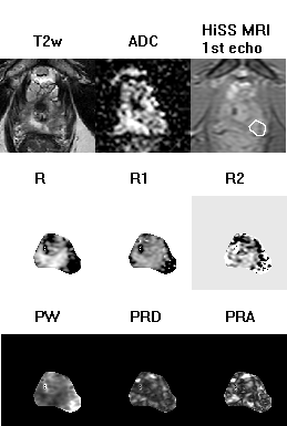
Figure 2:
The T2-weighted image, ADC map, shortest-TE HiSS MRI image, and HiSS
MRI-derived temporal domain (R, R1, R2) and spectral domain (PW, PRD, PRA) parameter
maps are shown here for a representative slice through the prostate of a 52-yo
man with a Gleason score 7 (3+4) lesion (white outline).
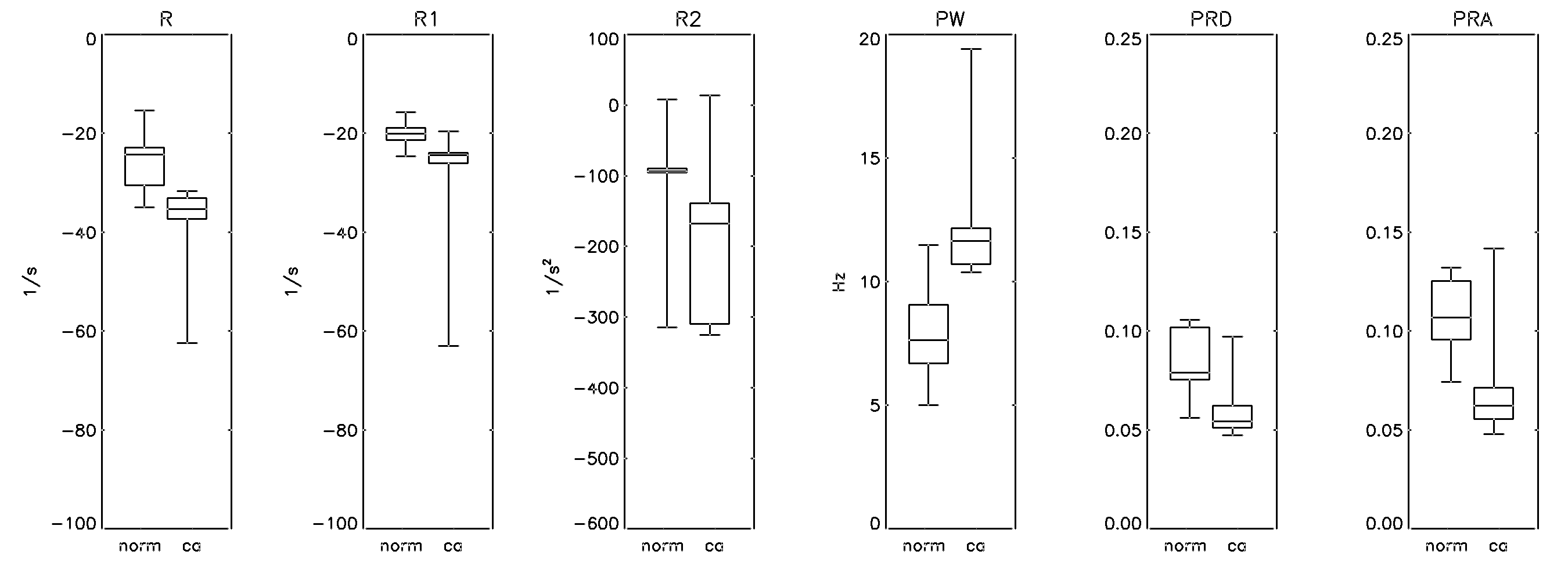
Figure 3:
Boxplots of HiSS MRI-derived parameters R, R1, R2, PW, PRD, and PRA are
shown, for normal and cancer ROIs. The
whiskers represent the full data range. All
HiSS-derived parameters were statistically significantly different (p <
0.05) between cancer and normal tissue ROIs, except R2.