Wenjun HU1, Ailian Liu1, Lihua Chen1, Zhiwei Shen2, Jiazheng Wang3, Yi Zhang4, and Qingwei Song1
1The First Affiliated Hospital of Dalian Medical University, Dalian, China, 2Philips Healthcare, Beijing, China, 3Philips Healthcare, Bejing, China, 4Zhejiang University, Hangzhou, China
1The First Affiliated Hospital of Dalian Medical University, Dalian, China, 2Philips Healthcare, Beijing, China, 3Philips Healthcare, Bejing, China, 4Zhejiang University, Hangzhou, China
Higher SNR and more clear anatomical
structure of APTw images in prostate was acquired with the
optimal scan parameters: the number of slices
of 7,
voxel
of 2×2 mm2, the
echo train length of 100, small FOV of 130×100×49mm3,
fold-over suppression of rest, SENSE of 1.1, phase
encoding direction of AP.
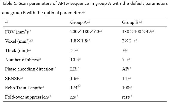
Table
1. Scan parameters of APTw sequence in group A with the
default parameters and group B with the
optimal parameters
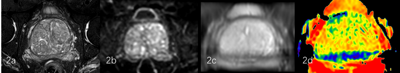
Figure2.
a 75-year-old male with BPH of group B. T2WI(2a), DWI (2b), M0 (2c) (central zone
SNR=71.22, peripheral zone SNR=22.12), APTw (2d) images are
shown.