Mohammed R. S. Sunoqrot1, Kirsten M. Selnæs1,2, Elise Sandsmark2, Sverre Langørgen2, Helena Bertilsson3,4, Tone F. Bathen1,2, and Mattijs Elschot1,2
1Department of Circulation and Medical Imaging, NTNU, Norwegian University of Science and Technolog, Trondheim, Norway, 2Department of Radiology and Nuclear Medicine, St. Olavs Hospital, Trondheim University Hospital, Trondheim, Norway, 3Department of Cancer Research and Molecular, NTNU, Norwegian University of Science and Technolog, Trondheim, Norway, 4Department of Urology, St. Olavs Hospital, Trondheim University Hospital, Trondheim, Norway
1Department of Circulation and Medical Imaging, NTNU, Norwegian University of Science and Technolog, Trondheim, Norway, 2Department of Radiology and Nuclear Medicine, St. Olavs Hospital, Trondheim University Hospital, Trondheim, Norway, 3Department of Cancer Research and Molecular, NTNU, Norwegian University of Science and Technolog, Trondheim, Norway, 4Department of Urology, St. Olavs Hospital, Trondheim University Hospital, Trondheim, Norway
The
repeatability of the best-performing deep learning-based prostate segmentation methods
is comparable to that of manual segmentations, which is important for clinical applications
based on multiple scans in time, such as active surveillance.
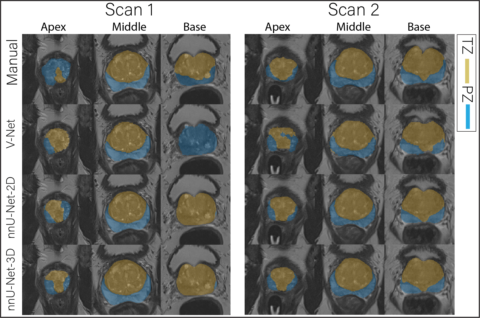
Figure 1 The
middle slice for the whole prostate, apex and base of a randomly selected case
segmented (peripheral zone (PZ) and transition zone (TZ)) by different
approaches for scan 1 and 2.
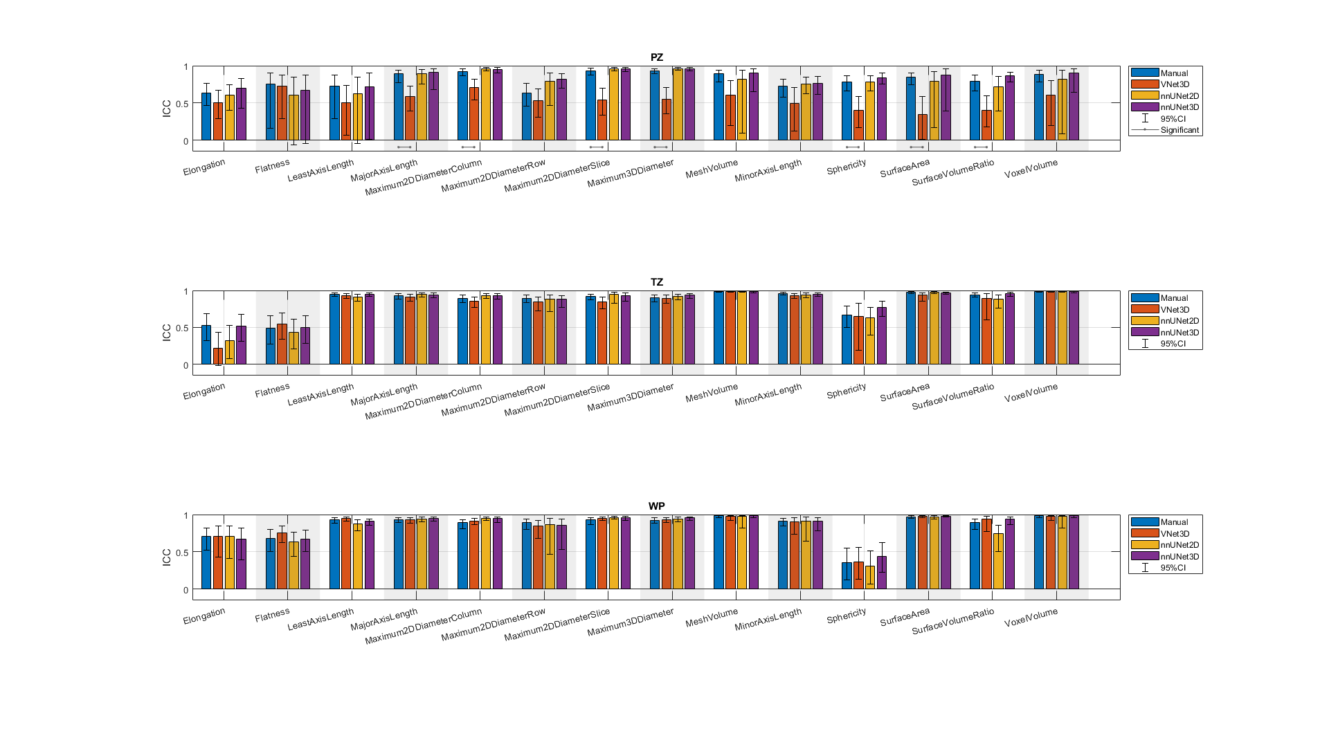
Figure 3 The single score intra-class correlation coefficient (ICC)
of the shape features extracted from the whole prostate gland (WP), peripheral
zone (PZ) and transition zone (TZ) for the
investigated methods. The methods connected with (-) were significantly
different. In both manual and DL-based segmentations, Elongation,
Flatness and Sphericity had a remarkably lower ICC than the other features in
WP and TZ.