Nader Aldoj1, Federico Biavati1, Sebastian Stober2, Marc Dewey1, Patrick Asbach1, and Ingolf Sack1
1Charité, Berlin, Germany, 2Ovgu Magdeburg, Magdeburg, Germany
1Charité, Berlin, Germany, 2Ovgu Magdeburg, Magdeburg, Germany
MR
elastography
maps can be used for prostate and zones segmentation due to the
excellent segmentation results even when compared to the standard
high-resolution T2w that is mostly used for anatomical segmentation.
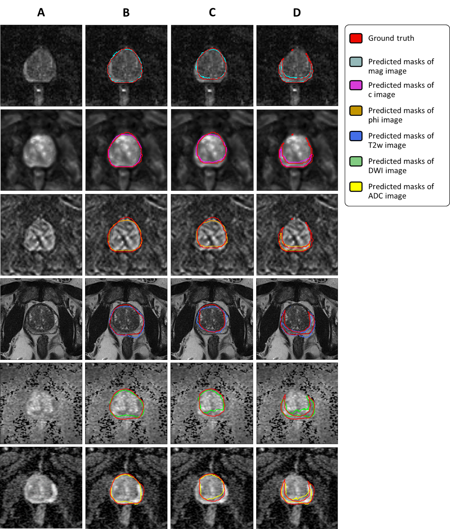
Figure 1: Segmentation
examples of individual models: first column (A) shows the
original image, second (B), third (C) and forth column (D) show masks
of prostate, central and peripheral zones, respectively. Rows from
top to bottom represents mag, c, phi maps, T2w, DWI, and ADC
respectively.
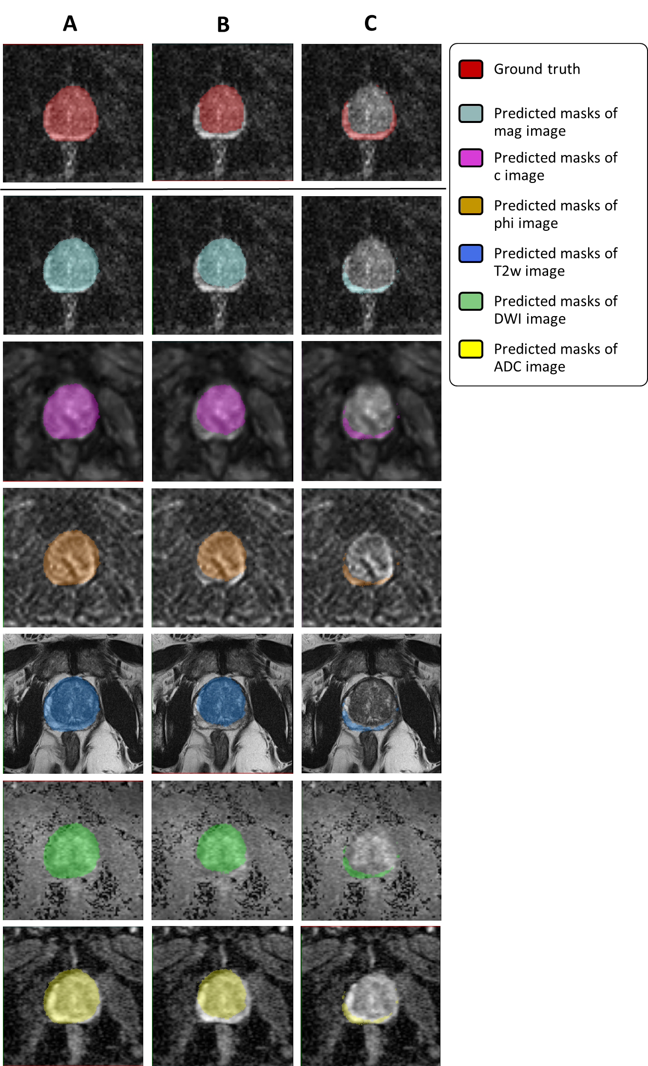
Figure 2: Examples
of segmentation results of the unified model: All segmented masks
resulted from combining all mre maps as input, and the resulting
masks were propagated to all other registered sequences. Columns A, B
and C show masks of the prostate, CZ and PZ respectively. Top row is
the mag images together with overlaid ground
truth
masks. The second row at the top to bottom show the predicted masks
overlaid on mag, c, phi, T2w, DWI and ADC images respectively.