Matthew Gibbons1, Jeffry P Simko2,3, Peter R Carroll2, and Susan Noworolski1
1Radiology and Biomedical Imaging, University of California, San Francisco, San Francisco, CA, United States, 2Urology, University of California, San Francisco, San Francisco, CA, United States, 3Pathology, University of California, San Francisco, San Francisco, CA, United States
1Radiology and Biomedical Imaging, University of California, San Francisco, San Francisco, CA, United States, 2Urology, University of California, San Francisco, San Francisco, CA, United States, 3Pathology, University of California, San Francisco, San Francisco, CA, United States
Multi-parametric MRI (mpMRI) is a clinically useful tool to assess prostate cancer. This study showed the feasibility of MRI generated cancer risk maps to detect cancer lesions >0.1cc and quantify the volume of cancer. The maps were validated by histopathology after prostatectomy.
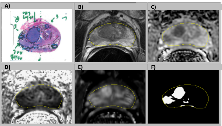
Fig 1: Information from a) histopathology, mpMRI images b) T2W, c) ADC, d) FA, and e) DCE) was combined in a logistic regression model to generate the f) cancer risk .aps in the TZ and the PZ. In this example, a combination in an ROI of hypointense T2W and ADC, hyperintense DCE resulted in a high-risk region in the cancer maps.
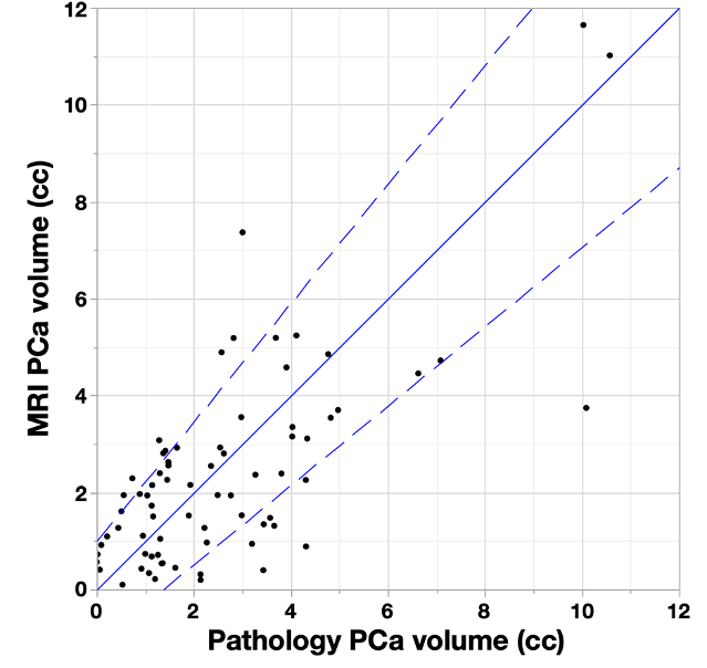
Fig. 2: Prostate cancer volume comparison of MRI cancer map vs histopathology. Cases with overestimated (underestimated) cancer volume are above (below) the solid blue one-to-one line. Dashed bounding lines were defined to indicate outliers (1cc ± 1.2·volume_pathology). 80% of cases were within the boundaries.