Patricia M Johnson1, Angela Tong1, Paul Smereka1, Awani Donthireddy1, Robert Petrocelli1, Hersh Chandarana1, and Florian Knoll1
1Center for Advanced Imaging Innovation and Research (CAI2R), Department of Radiology, New York University School of Medicine, New york, NY, United States
1Center for Advanced Imaging Innovation and Research (CAI2R), Department of Radiology, New York University School of Medicine, New york, NY, United States
Diagnostic quality highly accelerated axial and coronal T2-weighted prostate images were reconstructed using deep learning methods with ≤1 minute of acquisition time, which can enable rapid screening prostate MRI.
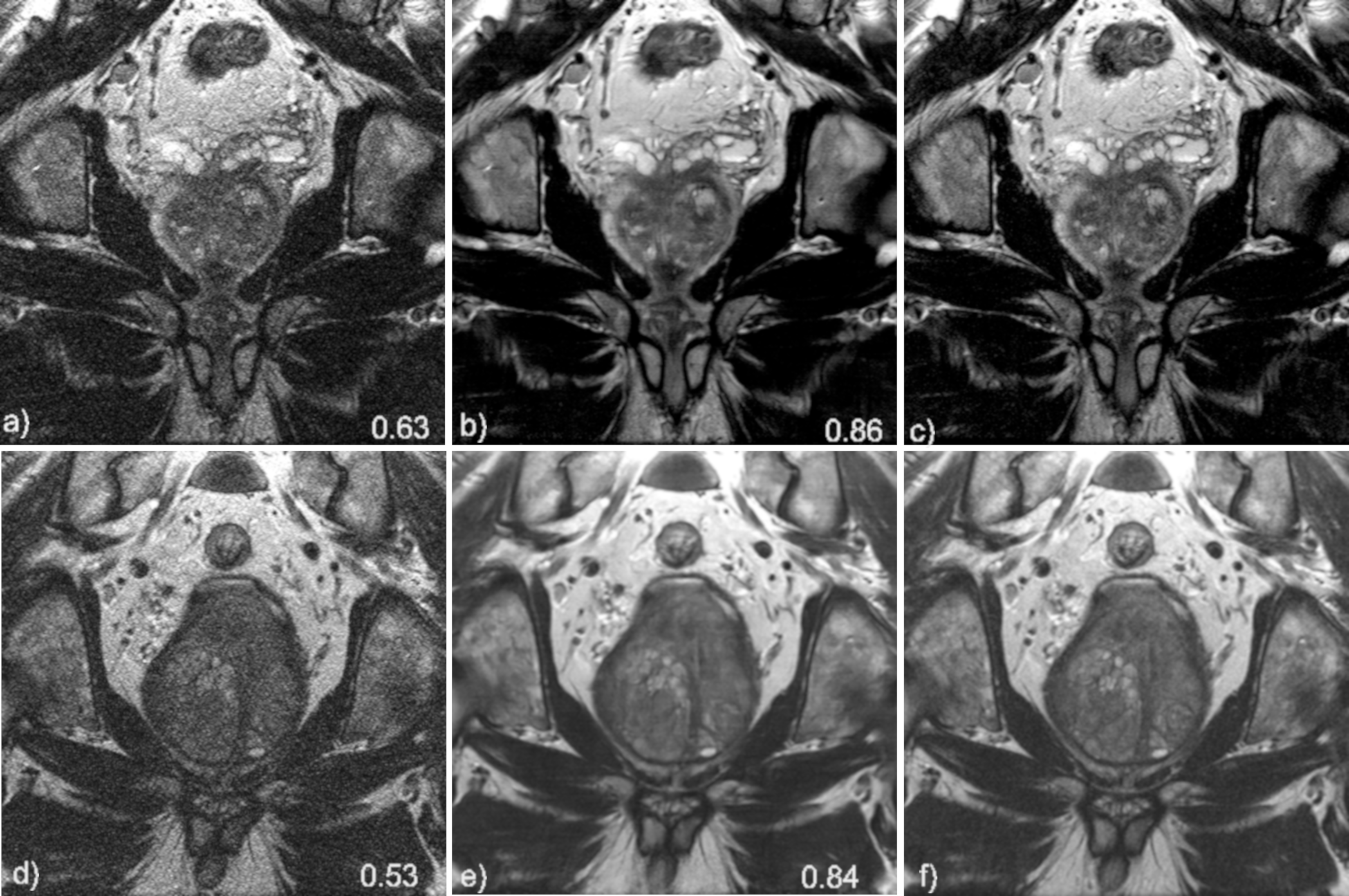
Figure 4. Coronal image results for subject 1 (top row) and subject 2 (bottom row). Soft-sense reconstructions are shown in a) and d). VN reconstructions are shown in b) and e) while the fully sampled, ground-truth images are shown in c) and f). The value indicated on the images is the calculated SSIM of the slice shown.
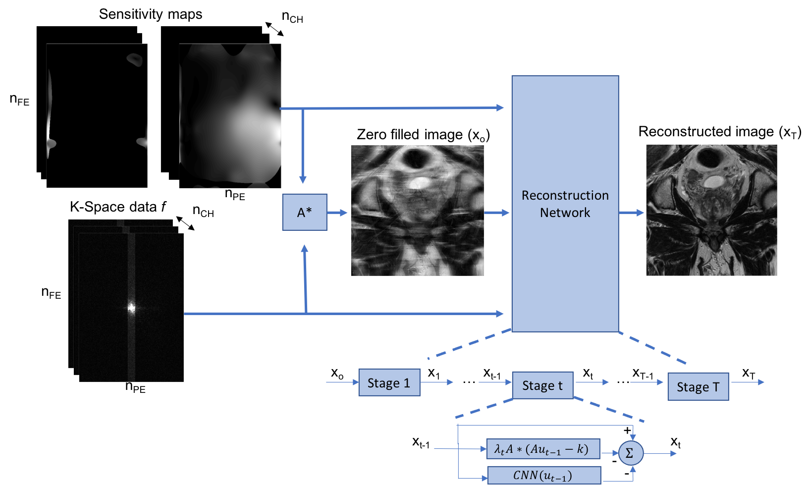
Figure 1. Data processing pipeline, and structure of the reconstruction network. First, a zero-filled reconstruction is generated from the under-sampled k space and two sets of coil sensitivity maps. Two sets of sensitivity estimates are required because the anatomy extends beyond the field of view, as described in Uecker et al.6 Then, this reconstruction, along with the raw k-space data and sensitivity maps are passed as the input to the reconstruction network. The reconstruction network has 10 stages (t =10), each stage applies data consistency and regularization.